 Movie
Movie Controller

[English] 日本語
 Yorodumi
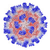
- PDB-8vgr: Cryo-EM structure of Tulane virus 9-6-17 variant capsid protein V... -
+ Open data

- Basic information
| Entry | Database: PDB / ID: 8vgr | ||||||
|---|---|---|---|---|---|---|---|
| Title | Cryo-EM structure of Tulane virus 9-6-17 variant capsid protein VP1 5-12-18 | ||||||
 Components Components | Capsid protein | ||||||
 Keywords Keywords | VIRUS / virion capsid / capsid protein / Tulane virus | ||||||
| Function / homology | Calicivirus coat protein C-terminal / Calicivirus coat protein C-terminal / Calicivirus coat protein / Calicivirus coat protein / Viral coat protein subunit / Capsid protein Function and homology information Function and homology information | ||||||
| Biological species |  Tulane virus Tulane virus | ||||||
| Method | ELECTRON MICROSCOPY / single particle reconstruction / cryo EM / Resolution: 3.2 Å | ||||||
 Authors Authors | Sun, C. / Jiang, W. | ||||||
| Funding support |  United States, 1items United States, 1items
| ||||||
 Citation Citation | Journal: Biomolecules / Year: 2024 Title: The 2.6 Å Structure of a Tulane Virus Variant with Minor Mutations Leading to Receptor Change. Authors: Chen Sun / Pengwei Huang / Xueyong Xu / Frank S Vago / Kunpeng Li / Thomas Klose / Xi Jason Jiang / Wen Jiang / Abstract: Human noroviruses (HuNoVs) are a major cause of acute gastroenteritis, contributing significantly to annual foodborne illness cases. However, studying these viruses has been challenging due to ...Human noroviruses (HuNoVs) are a major cause of acute gastroenteritis, contributing significantly to annual foodborne illness cases. However, studying these viruses has been challenging due to limitations in tissue culture techniques for over four decades. Tulane virus (TV) has emerged as a crucial surrogate for HuNoVs due to its close resemblance in amino acid composition and the availability of a robust cell culture system. Initially isolated from rhesus macaques in 2008, TV represents a novel belonging to the genus. Its significance lies in sharing the same host cell receptor, histo-blood group antigen (HBGA), as HuNoVs. In this study, we introduce, through cryo-electron microscopy (cryo-EM), the structure of a specific TV variant (the 9-6-17 TV) that has notably lost its ability to bind to its receptor, B-type HBGA-a finding confirmed using an enzyme-linked immunosorbent assay (ELISA). These results offer a profound insight into the genetic modifications occurring in TV that are necessary for adaptation to cell culture environments. This research significantly contributes to advancing our understanding of the genetic changes that are pivotal to successful adaptation, shedding light on fundamental aspects of evolution. | ||||||
| History |
|
- Structure visualization
| Structure viewer | Molecule: MolmilJmol/JSmol |
|---|
- Downloads & links
-Download
| PDBx/mmCIF format | 8vgr.cif.gz | 544 KB | Display | PDBx/mmCIF format |
|---|---|---|---|---|
| PDB format | pdb8vgr.ent.gz | 456.3 KB | Display | PDB format |
| PDBx/mmJSON format | 8vgr.json.gz | Tree view | PDBx/mmJSON format | |
| Others |  Other downloads Other downloads |
-Validation report
| Summary document | 8vgr_validation.pdf.gz | 1.5 MB | Display | wwPDB validaton report |
|---|---|---|---|---|
| Full document | 8vgr_full_validation.pdf.gz | 1.5 MB | Display | |
| Data in XML | 8vgr_validation.xml.gz | 59.5 KB | Display | |
| Data in CIF | 8vgr_validation.cif.gz | 86.1 KB | Display | |
| Arichive directory | https://data.pdbj.org/pub/pdb/validation_reports/vg/8vgrftp://data.pdbj.org/pub/pdb/validation_reports/vg/8vgr | HTTPS FTP |
-Related structure data
| Related structure data |  43222MC  8vjrC  8vjsC M: map data used to model this data C: citing same article ( |
|---|---|
| Similar structure data |
-Links
 PDBj
PDBj
- Assembly
| Deposited unit | 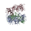
|
|---|---|
| 1 | x 60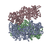
|
-Components
| #1: Protein | Mass: 57933.172 Da / Num. of mol.: 3 / Source method: isolated from a natural source / Source: (natural) Tulane virus / References: UniProt: B2Y6D0 |
|---|
-Experimental details
-Experiment
| Experiment | Method: ELECTRON MICROSCOPY |
|---|---|
| EM experiment | Aggregation state: PARTICLE / 3D reconstruction method: single particle reconstruction |
- Sample preparation
| Component | Name: Tulane virus / Type: VIRUS Details: The Tulane virus was purified from the LLC-MK2 cell line. Entity ID: all / Source: NATURAL |
|---|---|
| Molecular weight | Value: 57 MDa / Experimental value: NO |
| Source (natural) | Organism: Tulane virus / Strain: 9-6-17 |
| Details of virus | Empty: NO / Enveloped: NO / Isolate: STRAIN / Type: VIRION |
| Natural host | Organism: Macaca mulatta |
| Virus shell | Diameter: 400 nm |
| Buffer solution | pH: 7.4 / Details: PBS |
| Specimen | Embedding applied: NO / Shadowing applied: NO / Staining applied: NO / Vitrification applied: YES / Details: Purified virus |
| Specimen support | Details: Lacey grid coated with graphene oxide / Grid material: COPPER / Grid mesh size: 300 divisions/in. / Grid type: EMS Lacey Carbon |
| Vitrification | Instrument: GATAN CRYOPLUNGE 3 / Cryogen name: ETHANE / Humidity: 100 % / Chamber temperature: 293.15 K |
- Electron microscopy imaging
| Experimental equipment |  Model: Titan Krios / Image courtesy: FEI Company |
|---|---|
| Microscopy | Model: FEI TITAN KRIOS |
| Electron gun | Electron source:  FIELD EMISSION GUN / Accelerating voltage: 300 kV / Illumination mode: OTHER FIELD EMISSION GUN / Accelerating voltage: 300 kV / Illumination mode: OTHER |
| Electron lens | Mode: BRIGHT FIELD / Nominal magnification: 81000 X / Nominal defocus max: 2200 nm / Nominal defocus min: 800 nm |
| Image recording | Electron dose: 23 e/Å2 / Film or detector model: GATAN K2 SUMMIT (4k x 4k) / Num. of grids imaged: 1 / Num. of real images: 969 |
| Image scans | Movie frames/image: 35 |
- Processing
| EM software |
| |||||||||||||||||||||||||||||||||||||||
|---|---|---|---|---|---|---|---|---|---|---|---|---|---|---|---|---|---|---|---|---|---|---|---|---|---|---|---|---|---|---|---|---|---|---|---|---|---|---|---|---|
| CTF correction | Type: NONE | |||||||||||||||||||||||||||||||||||||||
| Symmetry | Point symmetry: I (icosahedral) | |||||||||||||||||||||||||||||||||||||||
| 3D reconstruction | Resolution: 3.2 Å / Resolution method: FSC 0.143 CUT-OFF / Num. of particles: 11645 / Symmetry type: POINT | |||||||||||||||||||||||||||||||||||||||
| Atomic model building | PDB-ID: 8VG6 8vg6 Accession code: 8VG6 / Source name: PDB / Type: experimental model | |||||||||||||||||||||||||||||||||||||||
| Refine LS restraints |
|
