 Movie
Movie Controller

[English] 日本語
 Yorodumi
- PDB-8sv0: The crystal structure of the classical binding interface of Impor... -
+ Open data

- Basic information
| Entry | Database: PDB / ID: 8sv0 | ||||||
|---|---|---|---|---|---|---|---|
| Title | The crystal structure of the classical binding interface of Importin alpha 2 and nuclear localisation signal sequence in Psittacine siadenovirus core protein VII | ||||||
 Components Components |
| ||||||
 Keywords Keywords | PROTEIN TRANSPORT/VIRAL PROTEIN /  Adenovirus protein VII / Nuclear localization / Importin alpha / DNA virus / PROTEIN TRANSPORT-VIRAL PROTEIN complex Adenovirus protein VII / Nuclear localization / Importin alpha / DNA virus / PROTEIN TRANSPORT-VIRAL PROTEIN complex | ||||||
| Function / homology |  Function and homology information Function and homology informationSensing of DNA Double Strand Breaks / entry of viral genome into host nucleus through nuclear pore complex via importin / positive regulation of viral life cycle / NLS-dependent protein nuclear import complex / postsynapse to nucleus signaling pathway / nuclear import signal receptor activity / nuclear localization sequence binding / NLS-bearing protein import into nucleus / host cell / cytoplasmic stress granule ...Sensing of DNA Double Strand Breaks / entry of viral genome into host nucleus through nuclear pore complex via importin / positive regulation of viral life cycle / NLS-dependent protein nuclear import complex / postsynapse to nucleus signaling pathway / nuclear import signal receptor activity / nuclear localization sequence binding / NLS-bearing protein import into nucleus / host cell / cytoplasmic stress granule / protein import into nucleus / DNA-binding transcription factor binding / postsynaptic density / glutamatergic synapse / nucleoplasm / nucleus / cytosolSimilarity search - Function | ||||||
| Biological species |  Mus musculus (house mouse) Mus musculus (house mouse) Psittacine siadenovirus F Psittacine siadenovirus F | ||||||
| Method | X-RAY DIFFRACTION / SYNCHROTRON / MOLECULAR REPLACEMENT / Resolution: 2.2 Å | ||||||
 Authors Authors | Athukorala, A. / Sarker, S. / Forwood, J.K. / Donnelly, C.M. | ||||||
| Funding support |  Australia, 1items Australia, 1items
| ||||||
 Citation Citation | Journal: To Be Published Title: Structural and functional characterization of nuclear localization signal of pVII protein of psittacine siadenovirus F demonstrates an independent transport of pVII protein into the nucleus Authors: Athukorala, A. / Sarker, S. / Forwood, J.K. / Donnelly, C.M. | ||||||
| History |
|
- Structure visualization
| Structure viewer | Molecule: MolmilJmol/JSmol |
|---|
- Downloads & links
-Download
| PDBx/mmCIF format | 8sv0.cif.gz | 176 KB | Display | PDBx/mmCIF format |
|---|---|---|---|---|
| PDB format | pdb8sv0.ent.gz | 138.2 KB | Display | PDB format |
| PDBx/mmJSON format | 8sv0.json.gz | Tree view | PDBx/mmJSON format | |
| Others |  Other downloads Other downloads |
-Validation report
| Arichive directory | https://data.pdbj.org/pub/pdb/validation_reports/sv/8sv0ftp://data.pdbj.org/pub/pdb/validation_reports/sv/8sv0 | HTTPS FTP |
|---|
-Related structure data
| Related structure data | 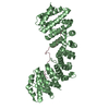 3ukxS S: Starting model for refinement |
|---|---|
| Similar structure data |
-Links
 PDBj
PDBj


- Assembly
| Deposited unit | 
| ||||||||
|---|---|---|---|---|---|---|---|---|---|
| 1 |
| ||||||||
| Unit cell |
|
-Components
| #1: Protein | / Importin alpha P1 / Karyopherin subunit alpha-2 / Pendulin / Pore targeting complex 58 kDa subunit ...Importin alpha P1 / Karyopherin subunit alpha-2 / Pendulin / Pore targeting complex 58 kDa subunit / PTAC58 / RAG cohort protein 1 / SRP1-alpha Mass: 55268.473 Da / Num. of mol.: 1 Source method: isolated from a genetically manipulated source Source: (gene. exp.) Mus musculus (house mouse) / Gene: Kpna2, Rch1 / Production host:  Escherichia coli BL21(DE3) (bacteria) / References: UniProt: P52293 Escherichia coli BL21(DE3) (bacteria) / References: UniProt: P52293 | ||||||
|---|---|---|---|---|---|---|---|
| #2: Protein/peptide | Mass: 1090.326 Da / Num. of mol.: 2 / Fragment: nuclear localization signal / Source method: obtained synthetically / Source: (synth.) Psittacine siadenovirus F#3: Chemical |   Mass: 22.990 Da / Num. of mol.: 2 / Source method: obtained synthetically / Formula: Na Mass: 22.990 Da / Num. of mol.: 2 / Source method: obtained synthetically / Formula: Na#4: Water | ChemComp-HOH / | Water Mass: 18.015 Da / Num. of mol.: 76 / Source method: isolated from a natural source / Formula: H2O Mass: 18.015 Da / Num. of mol.: 76 / Source method: isolated from a natural source / Formula: H2OHas ligand of interest | N | |
-Experimental details
-Experiment
| Experiment | Method: X-RAY DIFFRACTION / Number of used crystals: 1 |
|---|
- Sample preparation
| Crystal | Density Matthews: 2.87 Å3/Da / Density % sol: 57.21 % |
|---|---|
| Crystal grow | Temperature: 296 K / Method: vapor diffusion, hanging drop / pH: 6.5 Details: 0.6 M sodium citrate, 0.1 M sodium HEPES, 1% dithiothreitol |
-Data collection
| Diffraction | Mean temperature: 100 K / Serial crystal experiment: N |
|---|---|
| Diffraction source | Source: SYNCHROTRON / Site: Australian Synchrotron / Beamline: MX2 / Wavelength: 0.9537 Å |
| Detector | Type: DECTRIS EIGER X 16M / Detector: PIXEL / Date: Oct 5, 2022 |
| Radiation | Protocol: SINGLE WAVELENGTH / Monochromatic (M) / Laue (L): M / Scattering type: x-ray |
| Radiation wavelength | Wavelength: 0.9537 Å / Relative weight: 1 |
| Reflection | Resolution: 2.2→29.67 Å / Num. obs: 34168 / % possible obs: 99.7 % / Redundancy: 5.3 % / Biso Wilson estimate: 47.44 Å2 / CC1/2: 0.992 / Rmerge(I) obs: 0.163 / Rpim(I) all: 0.075 / Net I/σ(I): 4 |
| Reflection shell | Resolution: 2.2→2.27 Å / Rmerge(I) obs: 1.791 / Num. unique obs: 2903 / CC1/2: 0.319 / Rpim(I) all: 0.842 / % possible all: 99.5 |
- Processing
| Software |
| |||||||||||||||||||||||||||||||||||||||||||||||||||||||||||||||||||||||||||||||||||||||||||
|---|---|---|---|---|---|---|---|---|---|---|---|---|---|---|---|---|---|---|---|---|---|---|---|---|---|---|---|---|---|---|---|---|---|---|---|---|---|---|---|---|---|---|---|---|---|---|---|---|---|---|---|---|---|---|---|---|---|---|---|---|---|---|---|---|---|---|---|---|---|---|---|---|---|---|---|---|---|---|---|---|---|---|---|---|---|---|---|---|---|---|---|---|
| Refinement | Method to determine structure: MOLECULAR REPLACEMENT Starting model: PDB entry 3UKX Resolution: 2.2→29.67 Å / SU ML: 0.36 / Cross valid method: FREE R-VALUE / σ(F): 1.34 / Phase error: 27.62 / Stereochemistry target values: ML
| |||||||||||||||||||||||||||||||||||||||||||||||||||||||||||||||||||||||||||||||||||||||||||
| Solvent computation | Shrinkage radii: 0.9 Å / VDW probe radii: 1.1 Å / Solvent model: FLAT BULK SOLVENT MODEL | |||||||||||||||||||||||||||||||||||||||||||||||||||||||||||||||||||||||||||||||||||||||||||
| Displacement parameters | Biso mean: 69.82 Å2 | |||||||||||||||||||||||||||||||||||||||||||||||||||||||||||||||||||||||||||||||||||||||||||
| Refinement step | Cycle: LAST / Resolution: 2.2→29.67 Å
| |||||||||||||||||||||||||||||||||||||||||||||||||||||||||||||||||||||||||||||||||||||||||||
| Refine LS restraints |
| |||||||||||||||||||||||||||||||||||||||||||||||||||||||||||||||||||||||||||||||||||||||||||
| LS refinement shell |
|
