 Movie
Movie Controller

[English] 日本語
 Yorodumi
- PDB-8sts: Crystal Structure of HIV-1 Reverse Transcriptase (Y181C, V106A) v... -
+ Open data

- Basic information
| Entry | Database: PDB / ID: 8sts | ||||||
|---|---|---|---|---|---|---|---|
| Title | Crystal Structure of HIV-1 Reverse Transcriptase (Y181C, V106A) varient in Complex with 5-(2-(2-(2,4-dioxo-3,4-dihydropyrimidin-1(2H)-yl)ethoxy)-4-fluorophenoxy)-7-fluoro-2-naphthonitrile (JLJ636), a non-nucleoside inhibitor | ||||||
 Components Components |
| ||||||
 Keywords Keywords |  VIRAL PROTEIN / Hydrolase / REVERSE TRANSCRIPTASE / ANTIVIRAL / DRUG DESIGN / HIV-1 VIRAL PROTEIN / Hydrolase / REVERSE TRANSCRIPTASE / ANTIVIRAL / DRUG DESIGN / HIV-1 | ||||||
| Function / homology |  Function and homology informationHIV-1 retropepsin / : / retroviral ribonuclease H / exoribonuclease H / : / exoribonuclease H activity / host multivesicular body / DNA integration / RNA-directed DNA polymerase / viral genome integration into host DNA ...HIV-1 retropepsin / : / retroviral ribonuclease H / exoribonuclease H / : / exoribonuclease H activity / host multivesicular body / DNA integration / RNA-directed DNA polymerase / viral genome integration into host DNA / viral penetration into host nucleus / establishment of integrated proviral latency / RNA-directed DNA polymerase activity / Transferases; Transferring phosphorus-containing groups; Nucleotidyltransferases / RNA-DNA hybrid ribonuclease activity / viral nucleocapsid / DNA recombination / Hydrolases; Acting on ester bonds / DNA-directed DNA polymerase / aspartic-type endopeptidase activity / DNA-directed DNA polymerase activity / symbiont entry into host cell / symbiont-mediated suppression of host gene expression / lipid binding / host cell nucleus / host cell plasma membrane / virion membrane / structural molecule activity / proteolysis / DNA binding / RNA binding / zinc ion binding / membrane Function and homology informationHIV-1 retropepsin / : / retroviral ribonuclease H / exoribonuclease H / : / exoribonuclease H activity / host multivesicular body / DNA integration / RNA-directed DNA polymerase / viral genome integration into host DNA ...HIV-1 retropepsin / : / retroviral ribonuclease H / exoribonuclease H / : / exoribonuclease H activity / host multivesicular body / DNA integration / RNA-directed DNA polymerase / viral genome integration into host DNA / viral penetration into host nucleus / establishment of integrated proviral latency / RNA-directed DNA polymerase activity / Transferases; Transferring phosphorus-containing groups; Nucleotidyltransferases / RNA-DNA hybrid ribonuclease activity / viral nucleocapsid / DNA recombination / Hydrolases; Acting on ester bonds / DNA-directed DNA polymerase / aspartic-type endopeptidase activity / DNA-directed DNA polymerase activity / symbiont entry into host cell / symbiont-mediated suppression of host gene expression / lipid binding / host cell nucleus / host cell plasma membrane / virion membrane / structural molecule activity / proteolysis / DNA binding / RNA binding / zinc ion binding / membraneSimilarity search - Function | ||||||
| Biological species |  Human immunodeficiency virus type 1 BH10 Human immunodeficiency virus type 1 BH10 Human immunodeficiency virus 1 Human immunodeficiency virus 1 | ||||||
| Method | X-RAY DIFFRACTION / SYNCHROTRON / MOLECULAR REPLACEMENT / Resolution: 3.02 Å | ||||||
 Authors Authors | Hollander, K. / Jorgensen, W.L. / Anderson, K.S. | ||||||
| Funding support |  United States, 1items United States, 1items
| ||||||
 Citation Citation | Journal: Protein Sci. / Year: 2023 Title: Exploring novel HIV-1 reverse transcriptase inhibitors with drug-resistant mutants: A double mutant surprise. Authors: Hollander, K. / Chan, A.H. / Frey, K.M. / Hunker, O. / Ippolito, J.A. / Spasov, K.A. / Yeh, Y.J. / Jorgensen, W.L. / Ho, Y.C. / Anderson, K.S. | ||||||
| History |
|
- Structure visualization
| Structure viewer | Molecule: MolmilJmol/JSmol |
|---|
- Downloads & links
-Download
| PDBx/mmCIF format | 8sts.cif.gz | 382.2 KB | Display | PDBx/mmCIF format |
|---|---|---|---|---|
| PDB format | pdb8sts.ent.gz | 302.9 KB | Display | PDB format |
| PDBx/mmJSON format | 8sts.json.gz | Tree view | PDBx/mmJSON format | |
| Others |  Other downloads Other downloads |
-Validation report
| Arichive directory | https://data.pdbj.org/pub/pdb/validation_reports/st/8stsftp://data.pdbj.org/pub/pdb/validation_reports/st/8sts | HTTPS FTP |
|---|
-Related structure data
| Related structure data | 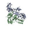 8stpC 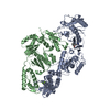 8stqC 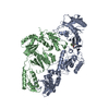 8strC 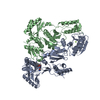 8sttC 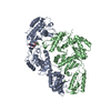 8stuC 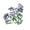 8stvC 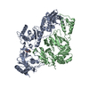 1eetS C: citing same article ( S: Starting model for refinement |
|---|---|
| Similar structure data |
-Links
 PDBj
PDBj


- Assembly
| Deposited unit | 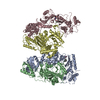
| ||||||||
|---|---|---|---|---|---|---|---|---|---|
| 1 | 
| ||||||||
| 2 | 
| ||||||||
| Unit cell |
|
-Components
| #1: Protein | Mass: 64014.309 Da / Num. of mol.: 2 / Mutation: Y181C, V106A, C280S, K172A, K173A Source method: isolated from a genetically manipulated source Source: (gene. exp.) Human immunodeficiency virus type 1 BH10Gene: gag-pol Production host:  Escherichia coli 'BL21-Gold(DE3)pLysS AG' (bacteria) Escherichia coli 'BL21-Gold(DE3)pLysS AG' (bacteria)References: UniProt: P03366, RNA-directed DNA polymerase, DNA-directed DNA polymerase, retroviral ribonuclease H#2: Protein | Mass: 50039.488 Da / Num. of mol.: 2 / Mutation: C280S Source method: isolated from a genetically manipulated source Source: (gene. exp.) Human immunodeficiency virus 1 / Gene: gag-polProduction host: Escherichia coli 'BL21-Gold(DE3)pLysS AG' (bacteria)References: UniProt: P03366 #3: Chemical | 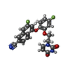  Mass: 435.380 Da / Num. of mol.: 2 / Source method: obtained synthetically / Formula: C23H15F2N3O4 / Feature type: SUBJECT OF INVESTIGATION Mass: 435.380 Da / Num. of mol.: 2 / Source method: obtained synthetically / Formula: C23H15F2N3O4 / Feature type: SUBJECT OF INVESTIGATION#4: Chemical |   Mass: 24.305 Da / Num. of mol.: 2 / Source method: obtained synthetically / Formula: Mg Mass: 24.305 Da / Num. of mol.: 2 / Source method: obtained synthetically / Formula: Mg#5: Water | ChemComp-HOH / | Water Mass: 18.015 Da / Num. of mol.: 12 / Source method: isolated from a natural source / Formula: H2O Mass: 18.015 Da / Num. of mol.: 12 / Source method: isolated from a natural source / Formula: H2OHas ligand of interest | Y | |
|---|
-Experimental details
-Experiment
| Experiment | Method: X-RAY DIFFRACTION / Number of used crystals: 1 |
|---|
- Sample preparation
| Crystal | Density Matthews: 3.24 Å3/Da / Density % sol: 62.06 % |
|---|---|
| Crystal grow | Temperature: 277 K / Method: vapor diffusion, hanging drop / pH: 7 Details: 50 mM HEPES pH 7.0, 20% PEG 8000, 100 mM ammonium sulfate, 15 mM magnesium sulfate, and 5 mM spermine PH range: 6.5-7.5 |
-Data collection
| Diffraction | Mean temperature: 100 K / Serial crystal experiment: N |
|---|---|
| Diffraction source | Source: SYNCHROTRON / Site: NSLS-II / Beamline: 17-ID-1 / Wavelength: 0.920105 Å |
| Detector | Type: DECTRIS EIGER X 9M / Detector: PIXEL / Date: Aug 9, 2022 |
| Radiation | Protocol: SINGLE WAVELENGTH / Monochromatic (M) / Laue (L): M / Scattering type: x-ray |
| Radiation wavelength | Wavelength: 0.920105 Å / Relative weight: 1 |
| Reflection | Resolution: 3.02→128.312 Å / Num. obs: 56957 / % possible obs: 99.6 % / Redundancy: 7.1 % / CC1/2: 0.998 / Rmerge(I) obs: 0.121 / Rpim(I) all: 0.049 / Rrim(I) all: 0.13 / Net I/σ(I): 10.3 |
| Reflection shell | Resolution: 3.02→3.072 Å / Rmerge(I) obs: 2.914 / Mean I/σ(I) obs: 0.8 / Num. unique obs: 2810 / CC1/2: 0.375 / Rpim(I) all: 1.15 / Rrim(I) all: 3.134 |
- Processing
| Software |
| |||||||||||||||||||||||||||||||||||||||||||||||||||||||||||||||||||||||||||||||||||||||||||||||||||||||||||||||||||||||||||||||||||||||||||||||||||
|---|---|---|---|---|---|---|---|---|---|---|---|---|---|---|---|---|---|---|---|---|---|---|---|---|---|---|---|---|---|---|---|---|---|---|---|---|---|---|---|---|---|---|---|---|---|---|---|---|---|---|---|---|---|---|---|---|---|---|---|---|---|---|---|---|---|---|---|---|---|---|---|---|---|---|---|---|---|---|---|---|---|---|---|---|---|---|---|---|---|---|---|---|---|---|---|---|---|---|---|---|---|---|---|---|---|---|---|---|---|---|---|---|---|---|---|---|---|---|---|---|---|---|---|---|---|---|---|---|---|---|---|---|---|---|---|---|---|---|---|---|---|---|---|---|---|---|---|---|
| Refinement | Method to determine structure: MOLECULAR REPLACEMENT Starting model: 1EET Resolution: 3.02→38.11 Å / SU ML: 0.59 / Cross valid method: FREE R-VALUE / σ(F): 1.33 / Phase error: 35.43 / Stereochemistry target values: ML
| |||||||||||||||||||||||||||||||||||||||||||||||||||||||||||||||||||||||||||||||||||||||||||||||||||||||||||||||||||||||||||||||||||||||||||||||||||
| Solvent computation | Shrinkage radii: 0.9 Å / VDW probe radii: 1.1 Å / Solvent model: FLAT BULK SOLVENT MODEL | |||||||||||||||||||||||||||||||||||||||||||||||||||||||||||||||||||||||||||||||||||||||||||||||||||||||||||||||||||||||||||||||||||||||||||||||||||
| Refinement step | Cycle: LAST / Resolution: 3.02→38.11 Å
| |||||||||||||||||||||||||||||||||||||||||||||||||||||||||||||||||||||||||||||||||||||||||||||||||||||||||||||||||||||||||||||||||||||||||||||||||||
| Refine LS restraints |
| |||||||||||||||||||||||||||||||||||||||||||||||||||||||||||||||||||||||||||||||||||||||||||||||||||||||||||||||||||||||||||||||||||||||||||||||||||
| LS refinement shell |
|
