 Movie
Movie Controller


+ Open data

- Basic information
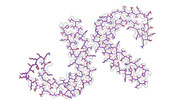
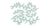
| Entry | Database: PDB / ID: 8seh | ||||||
|---|---|---|---|---|---|---|---|
| Title | PHF Tau from Down Syndrome | ||||||
 Components Components | Microtubule-associated protein tau Tau protein Tau protein | ||||||
 Keywords Keywords | NEUROPEPTIDE / PHF Tau / Tau Filament / Down Syndrome / Human Trisomy 21 | ||||||
| Function / homology |  Function and homology information Function and homology informationplus-end-directed organelle transport along microtubule / axonal transport / histone-dependent DNA binding / neurofibrillary tangle assembly / positive regulation of diacylglycerol kinase activity / negative regulation of establishment of protein localization to mitochondrion / neurofibrillary tangle / positive regulation of protein localization to synapse / microtubule lateral binding / tubulin complex ...plus-end-directed organelle transport along microtubule / axonal transport / histone-dependent DNA binding / neurofibrillary tangle assembly / positive regulation of diacylglycerol kinase activity / negative regulation of establishment of protein localization to mitochondrion / neurofibrillary tangle / positive regulation of protein localization to synapse / microtubule lateral binding / tubulin complex / phosphatidylinositol bisphosphate binding / main axon / regulation of long-term synaptic depression / negative regulation of kinase activity / negative regulation of tubulin deacetylation / generation of neurons / regulation of chromosome organization / positive regulation of protein localization / rRNA metabolic process / internal protein amino acid acetylation / regulation of mitochondrial fission / lipoprotein particle binding / intracellular distribution of mitochondria / axonal transport of mitochondrion / axon development / central nervous system neuron development / regulation of microtubule polymerization / microtubule polymerization / minor groove of adenine-thymine-rich DNA binding / negative regulation of mitochondrial membrane potential / dynactin binding / glial cell projection / apolipoprotein binding / protein polymerization / negative regulation of mitochondrial fission / axolemma / Caspase-mediated cleavage of cytoskeletal proteins / regulation of microtubule polymerization or depolymerization / positive regulation of axon extension / supramolecular fiber organization / Activation of AMPK downstream of NMDARs / regulation of microtubule cytoskeleton organization / stress granule assembly / cytoplasmic microtubule organization / regulation of cellular response to heat / regulation of calcium-mediated signaling / axon cytoplasm / positive regulation of microtubule polymerization / cellular response to brain-derived neurotrophic factor stimulus / somatodendritic compartment / synapse assembly / phosphatidylinositol binding / nuclear periphery / cellular response to nerve growth factor stimulus / positive regulation of superoxide anion generation / protein phosphatase 2A binding / regulation of autophagy / astrocyte activation / synapse organization / microglial cell activation / response to lead ion / Hsp90 protein binding / regulation of synaptic plasticity / PKR-mediated signaling / protein homooligomerization / cytoplasmic ribonucleoprotein granule / memory / microtubule cytoskeleton organization / cellular response to reactive oxygen species / SH3 domain binding / neuron projection development / activation of cysteine-type endopeptidase activity involved in apoptotic process / microtubule cytoskeleton / protein-macromolecule adaptor activity / single-stranded DNA binding / cell-cell signaling / cellular response to heat / cell body / actin binding / growth cone / protein-folding chaperone binding / double-stranded DNA binding / microtubule binding / microtubule / amyloid fibril formation / sequence-specific DNA binding / dendritic spine / learning or memory / neuron projection / nuclear speck / membrane raft / axon / negative regulation of gene expression / dendrite / neuronal cell body / DNA damage response / protein kinase binding / enzyme binding / mitochondrion / DNA bindingSimilarity search - Function | ||||||
| Biological species |  Homo sapiens (human) Homo sapiens (human) | ||||||
| Method | ELECTRON MICROSCOPY / helical reconstruction / cryo EM / Resolution: 2.9 Å | ||||||
 Authors Authors | Hoq, M.R. / Bharath, S.R. / Jiang, W. / Vago, F.S. | ||||||
| Funding support |  United States, 1items United States, 1items
| ||||||
 Citation Citation | Journal: Nat Struct Mol Biol / Year: 2024 Title: Cryo-EM structures of amyloid-β and tau filaments in Down syndrome. Authors: Anllely Fernandez / Md Rejaul Hoq / Grace I Hallinan / Daoyi Li / Sakshibeedu R Bharath / Frank S Vago / Xiaoqi Zhang / Kadir A Ozcan / Kathy L Newell / Holly J Garringer / Wen Jiang / ...Authors: Anllely Fernandez / Md Rejaul Hoq / Grace I Hallinan / Daoyi Li / Sakshibeedu R Bharath / Frank S Vago / Xiaoqi Zhang / Kadir A Ozcan / Kathy L Newell / Holly J Garringer / Wen Jiang / Bernardino Ghetti / Ruben Vidal / Abstract: Adult individuals with Down syndrome (DS) develop Alzheimer disease (AD). Whether there is a difference between AD in DS and AD regarding the structure of amyloid-β (Aβ) and tau filaments is ...Adult individuals with Down syndrome (DS) develop Alzheimer disease (AD). Whether there is a difference between AD in DS and AD regarding the structure of amyloid-β (Aβ) and tau filaments is unknown. Here we report the structure of Aβ and tau filaments from two DS brains. We found two Aβ filaments (types IIIa and IIIb) that differ from those previously reported in sporadic AD and two types of Aβ filaments (I and II) identical to those found in sporadic and familial AD. Tau filaments (paired helical filaments and straight filaments) were identical to those in AD, supporting the notion of a common mechanism through which amyloids trigger aggregation of tau. This knowledge is important for understanding AD in DS and assessing whether adults with DS could be included in AD clinical trials. | ||||||
| History |
|
- Structure visualization
| Structure viewer | Molecule: MolmilJmol/JSmol |
|---|
- Downloads & links
-Download
| PDBx/mmCIF format | 8seh.cif.gz | 138.2 KB | Display | PDBx/mmCIF format |
|---|---|---|---|---|
| PDB format | pdb8seh.ent.gz | 111.6 KB | Display | PDB format |
| PDBx/mmJSON format | 8seh.json.gz | Tree view | PDBx/mmJSON format | |
| Others |  Other downloads Other downloads |
-Validation report
| Arichive directory | https://data.pdbj.org/pub/pdb/validation_reports/se/8sehftp://data.pdbj.org/pub/pdb/validation_reports/se/8seh | HTTPS FTP |
|---|
-Related structure data
| Related structure data | 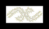 40411MC  8seiC  8sejC  8sekC  8selC M: map data used to model this data C: citing same article ( |
|---|---|
| Similar structure data |
-Links
 PDBj
PDBj


- Assembly
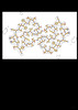
| Deposited unit | 
|
|---|---|
| 1 |
|
-Components
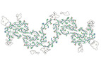
| #1: Protein | Tau protein / Neurofibrillary tangle protein / Paired helical filament-tau / PHF-tau Mass: 7940.141 Da / Num. of mol.: 10 Source method: isolated from a genetically manipulated source Source: (gene. exp.) Homo sapiens (human) / Gene: MAPT, MAPTL, MTBT1, TAU / Production host: Homo sapiens (human) / References: UniProt: P10636 |
|---|
-Experimental details
-Experiment
| Experiment | Method: ELECTRON MICROSCOPY |
|---|---|
| EM experiment | Aggregation state: FILAMENT / 3D reconstruction method: helical reconstruction |
- Sample preparation
| Component | Name: Helical Filaments / Type: TISSUE / Entity ID: all / Source: NATURAL |
|---|---|
| Source (natural) | Organism: Homo sapiens (human) |
| Buffer solution | pH: 7.2 |
| Specimen | Conc.: 1 mg/ml / Embedding applied: NO / Shadowing applied: NO / Staining applied: NO / Vitrification applied: YES |
| Vitrification | Cryogen name: ETHANE |
- Electron microscopy imaging
| Experimental equipment |  Model: Titan Krios / Image courtesy: FEI Company |
|---|---|
| Microscopy | Model: TFS KRIOS |
| Electron gun | Electron source: FIELD EMISSION GUN / Accelerating voltage: 300 kV / Illumination mode: SPOT SCAN |
| Electron lens | Mode: BRIGHT FIELDBright-field microscopy / Nominal magnification: 81000 X / Nominal defocus max: 5000 nm / Nominal defocus min: 500 nm / Cs: 2.7 mm / C2 aperture diameter: 100 µm |
| Specimen holder | Cryogen: NITROGEN |
| Image recording | Average exposure time: 1.103 sec. / Electron dose: 50.46 e/Å2 / Film or detector model: GATAN K3 (6k x 4k) |
- Processing
| EM software | Name: CTFFIND / Category: CTF correction |
|---|---|
| CTF correction | Type: PHASE FLIPPING AND AMPLITUDE CORRECTION |
| Helical symmerty | Angular rotation/subunit: 179.46 ° / Axial rise/subunit: 2.38 Å / Axial symmetry: C1 |
| 3D reconstruction | Resolution: 2.9 Å / Resolution method: FSC 0.143 CUT-OFF / Num. of particles: 90384 / Symmetry type: HELICAL |
