 Movie
Movie Controller


+ Open data

- Basic information
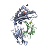
| Entry | Database: PDB / ID: 8sbl | |||||||||
|---|---|---|---|---|---|---|---|---|---|---|
| Title | Structure of HLA-A*24:02 in complex with peptide, LYLPVRVLI | |||||||||
 Components Components |
| |||||||||
 Keywords Keywords |  IMMUNE SYSTEM / Major Histocompatibility Complex (MHC) IMMUNE SYSTEM / Major Histocompatibility Complex (MHC) | |||||||||
| Function / homology |  Function and homology information Function and homology informationpositive regulation of ferrous iron binding / positive regulation of transferrin receptor binding / positive regulation of receptor binding / early endosome lumen / Nef mediated downregulation of MHC class I complex cell surface expression / DAP12 interactions / negative regulation of receptor binding / Endosomal/Vacuolar pathway / Antigen Presentation: Folding, assembly and peptide loading of class I MHC / cellular response to iron(III) ion ...positive regulation of ferrous iron binding / positive regulation of transferrin receptor binding / positive regulation of receptor binding / early endosome lumen / Nef mediated downregulation of MHC class I complex cell surface expression / DAP12 interactions / negative regulation of receptor binding / Endosomal/Vacuolar pathway / Antigen Presentation: Folding, assembly and peptide loading of class I MHC / cellular response to iron(III) ion / antigen processing and presentation of exogenous protein antigen via MHC class Ib, TAP-dependent / negative regulation of forebrain neuron differentiation / ER to Golgi transport vesicle membrane / regulation of erythrocyte differentiation / peptide antigen assembly with MHC class I protein complex / response to molecule of bacterial origin / regulation of iron ion transport / MHC class I peptide loading complex / HFE-transferrin receptor complex / T cell mediated cytotoxicity / cellular response to iron ion / antigen processing and presentation of endogenous peptide antigen via MHC class I / positive regulation of T cell cytokine production / MHC class I protein complex / multicellular organismal-level iron ion homeostasis / negative regulation of neurogenesis / peptide antigen assembly with MHC class II protein complex / positive regulation of receptor-mediated endocytosis / positive regulation of T cell mediated cytotoxicity / MHC class II protein complex / cellular response to nicotine / recycling endosome membrane / specific granule lumen / phagocytic vesicle membrane / peptide antigen binding / positive regulation of cellular senescence / antigen processing and presentation of exogenous peptide antigen via MHC class II / negative regulation of epithelial cell proliferation / Immunoregulatory interactions between a Lymphoid and a non-Lymphoid cell / Interferon gamma signaling / positive regulation of immune response / Modulation by Mtb of host immune system / sensory perception of smell / positive regulation of T cell activation / positive regulation of protein binding / tertiary granule lumen / DAP12 signaling / negative regulation of neuron projection development / MHC class II protein complex binding / late endosome membrane / T cell differentiation in thymus / ER-Phagosome pathway / iron ion transport / early endosome membrane / protein refolding / protein homotetramerization / intracellular iron ion homeostasis / amyloid fibril formation / learning or memory / Amyloid fiber formation / lysosomal membrane / external side of plasma membrane / endoplasmic reticulum lumen / Golgi membrane / focal adhesion / Neutrophil degranulation / SARS-CoV-2 activates/modulates innate and adaptive immune responses / structural molecule activity / Golgi apparatus / endoplasmic reticulum / protein homodimerization activity / extracellular space / extracellular exosome / extracellular region / membrane / identical protein binding / plasma membrane / cytosolSimilarity search - Function | |||||||||
| Biological species |  Homo sapiens (human) Homo sapiens (human) | |||||||||
| Method | X-RAY DIFFRACTION / SYNCHROTRON / MOLECULAR REPLACEMENT / Resolution: 3 Å | |||||||||
 Authors Authors | Mallik, L. / Young, M.C. / Sgourakis, N.G. | |||||||||
| Funding support |  United States, 2items United States, 2items
| |||||||||
 Citation Citation | Journal: Sci Immunol / Year: 2023 Title: Structural principles of peptide-centric chimeric antigen receptor recognition guide therapeutic expansion. Authors: Sun, Y. / Florio, T.J. / Gupta, S. / Young, M.C. / Marshall, Q.F. / Garfinkle, S.E. / Papadaki, G.F. / Truong, H.V. / Mycek, E. / Li, P. / Farrel, A. / Church, N.L. / Jabar, S. / Beasley, M. ...Authors: Sun, Y. / Florio, T.J. / Gupta, S. / Young, M.C. / Marshall, Q.F. / Garfinkle, S.E. / Papadaki, G.F. / Truong, H.V. / Mycek, E. / Li, P. / Farrel, A. / Church, N.L. / Jabar, S. / Beasley, M.D. / Kiefel, B.R. / Yarmarkovich, M. / Mallik, L. / Maris, J.M. / Sgourakis, N.G. #1: Journal: Sci Immunol / Year: 2023 Title: The chilling origin of germinal centers. Authors: Boehm, T. | |||||||||
| History |
|
- Structure visualization
| Structure viewer | Molecule: MolmilJmol/JSmol |
|---|
- Downloads & links
-Download
| PDBx/mmCIF format | 8sbl.cif.gz | 312.2 KB | Display | PDBx/mmCIF format |
|---|---|---|---|---|
| PDB format | pdb8sbl.ent.gz | 253.8 KB | Display | PDB format |
| PDBx/mmJSON format | 8sbl.json.gz | Tree view | PDBx/mmJSON format | |
| Others |  Other downloads Other downloads |
-Validation report
| Arichive directory | https://data.pdbj.org/pub/pdb/validation_reports/sb/8sblftp://data.pdbj.org/pub/pdb/validation_reports/sb/8sbl | HTTPS FTP |
|---|
-Related structure data

-Links
 PDBj
PDBj

- Assembly
| Deposited unit | 
| ||||||||
|---|---|---|---|---|---|---|---|---|---|
| 1 |
| ||||||||
| 2 |
| ||||||||
| 3 |
| ||||||||
| 4 |
| ||||||||
| Unit cell |
|
-Components
| #1: Protein | Mass: 32466.869 Da / Num. of mol.: 4 Source method: isolated from a genetically manipulated source Source: (gene. exp.) Homo sapiens (human) / Gene: HLA-A / Production host:  Escherichia coli (E. coli) / References: UniProt: A0A411J078 Escherichia coli (E. coli) / References: UniProt: A0A411J078#2: Protein | Beta-2 microglobulinMass: 11879.356 Da / Num. of mol.: 4 / Fragment: UNP residues 21-119 Source method: isolated from a genetically manipulated source Source: (gene. exp.) Homo sapiens (human) / Gene: B2M, CDABP0092, HDCMA22P / Production host: Escherichia coli (E. coli) / References: UniProt: P61769#3: Protein/peptide | Mass: 1086.390 Da / Num. of mol.: 4 / Source method: obtained synthetically / Source: (synth.) Homo sapiens (human) |
|---|
-Experimental details
-Experiment
| Experiment | Method: X-RAY DIFFRACTION / Number of used crystals: 1 |
|---|
- Sample preparation
| Crystal | Density Matthews: 2.45 Å3/Da / Density % sol: 49.84 % |
|---|---|
| Crystal grow | Temperature: 294 K / Method: vapor diffusion, hanging drop / pH: 6.5 Details: 0.05M MES monohydrate pH 6.5, 22.5% Polyethylene Glycol w/v 3350 |
-Data collection
| Diffraction | Mean temperature: 100 K / Serial crystal experiment: N |
|---|---|
| Diffraction source | Source: SYNCHROTRON / Site: NSLS-II / Beamline: 17-ID-1 / Wavelength: 0.92 Å |
| Detector | Type: DECTRIS EIGER X 9M / Detector: PIXEL / Date: Jan 27, 2023 |
| Radiation | Protocol: SINGLE WAVELENGTH / Monochromatic (M) / Laue (L): M / Scattering type: x-ray |
| Radiation wavelength | Wavelength: 0.92 Å / Relative weight: 1 |
| Reflection | Resolution: 3→105.19 Å / Num. obs: 34967 / % possible obs: 99.8 % / Redundancy: 3.5 % / Biso Wilson estimate: 36.83 Å2 / CC1/2: 0.877 / R split: 0.341 / Rmerge(I) obs: 0.388 / Rpim(I) all: 0.244 / Rrim(I) all: 0.459 / Χ2: 0.46 / Net I/σ(I): 2.8 |
| Reflection shell | Resolution: 3.05→3.21 Å / Redundancy: 3.5 % / Rmerge(I) obs: 1.203 / Mean I/σ(I) obs: 0.9 / Num. unique obs: 4832 / CC1/2: 0.419 / Rpim(I) all: 0.746 / Rrim(I) all: 1.418 / Χ2: 0.47 / % possible all: 100 |
- Processing
| Software |
| |||||||||||||||||||||||||||||||||||||||||||||||||||||||||||||||||||||||||||||||||||||||||||||||||||||||||
|---|---|---|---|---|---|---|---|---|---|---|---|---|---|---|---|---|---|---|---|---|---|---|---|---|---|---|---|---|---|---|---|---|---|---|---|---|---|---|---|---|---|---|---|---|---|---|---|---|---|---|---|---|---|---|---|---|---|---|---|---|---|---|---|---|---|---|---|---|---|---|---|---|---|---|---|---|---|---|---|---|---|---|---|---|---|---|---|---|---|---|---|---|---|---|---|---|---|---|---|---|---|---|---|---|---|---|
| Refinement | Method to determine structure: MOLECULAR REPLACEMENT / Resolution: 3→105.19 Å / SU ML: 0.49 / Cross valid method: FREE R-VALUE / σ(F): 1.34 / Phase error: 25.54 / Stereochemistry target values: ML
| |||||||||||||||||||||||||||||||||||||||||||||||||||||||||||||||||||||||||||||||||||||||||||||||||||||||||
| Solvent computation | Shrinkage radii: 0.9 Å / VDW probe radii: 1.1 Å / Solvent model: FLAT BULK SOLVENT MODEL | |||||||||||||||||||||||||||||||||||||||||||||||||||||||||||||||||||||||||||||||||||||||||||||||||||||||||
| Refinement step | Cycle: LAST / Resolution: 3→105.19 Å
| |||||||||||||||||||||||||||||||||||||||||||||||||||||||||||||||||||||||||||||||||||||||||||||||||||||||||
| Refine LS restraints |
| |||||||||||||||||||||||||||||||||||||||||||||||||||||||||||||||||||||||||||||||||||||||||||||||||||||||||
| LS refinement shell |
|
