 Movie
Movie Controller

[English] 日本語
 Yorodumi
- PDB-8r5i: In situ structure of the Vaccinia virus (WR) A4/A10 palisade trim... -
+ Open data

- Basic information
| Entry | Database: PDB / ID: 8r5i | ||||||
|---|---|---|---|---|---|---|---|
| Title | In situ structure of the Vaccinia virus (WR) A4/A10 palisade trimer in mature virions by flexible fitting into a cryoET map | ||||||
 Components Components |
| ||||||
 Keywords Keywords |  VIRAL PROTEIN / Palisade / A10 / A4 / p4a / Vaccinia VIRAL PROTEIN / Palisade / A10 / A4 / p4a / Vaccinia | ||||||
| Function / homology |  Function and homology informationvirion component / host cell endoplasmic reticulum-Golgi intermediate compartment membrane / structural molecule activity / membrane Function and homology informationvirion component / host cell endoplasmic reticulum-Golgi intermediate compartment membrane / structural molecule activity / membraneSimilarity search - Function | ||||||
| Biological species |  Vaccinia virus WR Vaccinia virus WR | ||||||
| Method | ELECTRON MICROSCOPY / subtomogram averaging / cryo EM / Resolution: 9.7 Å | ||||||
 Authors Authors | Calcraft, T. / Hernandez-Gonzalez, M. / Nans, A. / Rosenthal, P.B. / Way, M. | ||||||
| Funding support |  United Kingdom, 1items United Kingdom, 1items
| ||||||
 Citation Citation | Journal: mBio / Year: 2024 Title: Palisade structure in intact vaccinia virions. Authors: Miguel Hernandez-Gonzalez / Thomas Calcraft / Andrea Nans / Peter B Rosenthal / Michael Way / Abstract: Vaccinia virus assembly in the cytoplasm of infected cells involves the formation of a biconcave viral core inside the maturing viral particle. The boundary of the core is defined by a ...Vaccinia virus assembly in the cytoplasm of infected cells involves the formation of a biconcave viral core inside the maturing viral particle. The boundary of the core is defined by a pseudohexagonal palisade layer, composed of trimers projecting from an inner wall. To understand the assembly of this complex core architecture, we obtained a subnanometer structure of the palisade trimer by cryo-electron tomography and subtomogram averaging of purified intact virions. Using AlphaFold2 structure predictions, we determined that the palisade is formed from trimers of the proteolytically processed form of the viral protein A10. In addition, we found that each A10 protomer associates with an α-helix (residues 24-66) of A4. Cellular localization assays outside the context of infection demonstrate that the A4 N-terminus is necessary and sufficient to interact with A10. The interaction between A4 and A10 provides insights into how the palisade layer might become tightly associated with the viral membrane during virion maturation. Reconstruction of the palisade layer reveals that, despite local hexagonal ordering, the A10/A4 trimers are widely spaced, suggesting that additional components organize the lattice. This spacing would, however, allow the adoption of the characteristic biconcave shape of the viral core. Finally, we also found that the palisade incorporates multiple copies of a hexameric portal structure. We suggest that these portals are formed by E6, a viral protein that is essential for virion assembly and required to release viral mRNA from the core early in infection.IMPORTANCEPoxviruses such as variola virus (smallpox) and monkeypox cause diseases in humans. Other poxviruses, including vaccinia and modified vaccinia Ankara, are used as vaccine vectors. Given their importance, a greater structural understanding of poxvirus virions is needed. We now performed cryo-electron tomography of purified intact vaccinia virions to study the structure of the palisade, a protein lattice that defines the viral core boundary. We identified the main viral proteins that form the palisade and their interaction surfaces and provided new insights into the organization of the viral core. | ||||||
| History |
|
- Structure visualization
| Structure viewer | Molecule: MolmilJmol/JSmol |
|---|
- Downloads & links
-Download
| PDBx/mmCIF format | 8r5i.cif.gz | 398.3 KB | Display | PDBx/mmCIF format |
|---|---|---|---|---|
| PDB format | pdb8r5i.ent.gz | 321.1 KB | Display | PDB format |
| PDBx/mmJSON format | 8r5i.json.gz | Tree view | PDBx/mmJSON format | |
| Others |  Other downloads Other downloads |
-Validation report
| Arichive directory | https://data.pdbj.org/pub/pdb/validation_reports/r5/8r5iftp://data.pdbj.org/pub/pdb/validation_reports/r5/8r5i | HTTPS FTP |
|---|
-Related structure data
| Related structure data |  18918MC M: map data used to model this data C: citing same article ( |
|---|---|
| Similar structure data |


-Links
 PDBj
PDBj- Assembly
| Deposited unit | 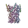
| ||||||||||||||||||||||||||||||||||||||||||||||||||||||||||||||||||||||||||||||||||||||||||||||||||||||||||||||||||||||||||||||||||||||||||||||||||||||||||||||||||||||||||||||||||||||||||||||||||||||||||
|---|---|---|---|---|---|---|---|---|---|---|---|---|---|---|---|---|---|---|---|---|---|---|---|---|---|---|---|---|---|---|---|---|---|---|---|---|---|---|---|---|---|---|---|---|---|---|---|---|---|---|---|---|---|---|---|---|---|---|---|---|---|---|---|---|---|---|---|---|---|---|---|---|---|---|---|---|---|---|---|---|---|---|---|---|---|---|---|---|---|---|---|---|---|---|---|---|---|---|---|---|---|---|---|---|---|---|---|---|---|---|---|---|---|---|---|---|---|---|---|---|---|---|---|---|---|---|---|---|---|---|---|---|---|---|---|---|---|---|---|---|---|---|---|---|---|---|---|---|---|---|---|---|---|---|---|---|---|---|---|---|---|---|---|---|---|---|---|---|---|---|---|---|---|---|---|---|---|---|---|---|---|---|---|---|---|---|---|---|---|---|---|---|---|---|---|---|---|---|---|---|---|---|---|
| 1 |
| ||||||||||||||||||||||||||||||||||||||||||||||||||||||||||||||||||||||||||||||||||||||||||||||||||||||||||||||||||||||||||||||||||||||||||||||||||||||||||||||||||||||||||||||||||||||||||||||||||||||||||
| Noncrystallographic symmetry (NCS) | NCS domain:
NCS domain segments: Auth asym-ID: A / Label asym-ID: A
NCS ensembles :
NCS oper:
|
-Components
| #1: Protein | Mass: 71073.477 Da / Num. of mol.: 3 Source method: isolated from a genetically manipulated source Source: (gene. exp.) Vaccinia virus WR / Gene: OPG136, VACWR129, A10L / Production host:  Homo sapiens (human) / References: UniProt: P16715 Homo sapiens (human) / References: UniProt: P16715#2: Protein | Mass: 30952.350 Da / Num. of mol.: 3 Source method: isolated from a genetically manipulated source Source: (gene. exp.) Vaccinia virus WR / Gene: OPG130, VACWR123, A4L / Production host: Homo sapiens (human) / References: UniProt: P29191 |
|---|
-Experimental details
-Experiment
| Experiment | Method: ELECTRON MICROSCOPY |
|---|---|
| EM experiment | Aggregation state: PARTICLE / 3D reconstruction method: subtomogram averaging |
- Sample preparation
| Component | Name: Vaccinia virus WR / Type: VIRUS / Details: A36-YdF / Entity ID: all / Source: RECOMBINANT |
|---|---|
| Molecular weight | Experimental value: NO |
| Source (natural) | Organism: Vaccinia virus WR |
| Source (recombinant) | Organism: Homo sapiens (human) |
| Details of virus | Empty: NO / Enveloped: YES / Isolate: STRAIN / Type: VIRION |
| Natural host | Organism: Homo sapiens |
| Buffer solution | pH: 7.5 |
| Specimen | Embedding applied: NO / Shadowing applied: NO / Staining applied: NO / Vitrification applied: YES |
| Specimen support | Details: 45mA / Grid material: COPPER / Grid mesh size: 300 divisions/in. / Grid type: Quantifoil R2/2 |
| Vitrification | Instrument: FEI VITROBOT MARK IV / Cryogen name: ETHANE / Humidity: 95 % / Chamber temperature: 295 K |
- Electron microscopy imaging
| Experimental equipment |  Model: Titan Krios / Image courtesy: FEI Company |
|---|---|
| Microscopy | Model: FEI TITAN KRIOS |
| Electron gun | Electron source: FIELD EMISSION GUN / Accelerating voltage: 300 kV / Illumination mode: FLOOD BEAM |
| Electron lens | Mode: BRIGHT FIELDBright-field microscopy / Nominal defocus max: 5000 nm / Nominal defocus min: 2000 nm / Cs: 2.7 mm / Alignment procedure: ZEMLIN TABLEAU |
| Specimen holder | Cryogen: NITROGEN / Specimen holder model: FEI TITAN KRIOS AUTOGRID HOLDER |
| Image recording | Electron dose: 2.4 e/Å2 / Avg electron dose per subtomogram: 98.6 e/Å2 / Film or detector model: TFS FALCON 4i (4k x 4k) |
| EM imaging optics | Energyfilter name: TFS Selectris / Energyfilter slit width: 10 eV |
- Processing
| EM software |
| ||||||||||||||||||||||||||||||||||||||||||||||||||||||||||||||||||||||||||||||||||||||||||||||||||||||||||
|---|---|---|---|---|---|---|---|---|---|---|---|---|---|---|---|---|---|---|---|---|---|---|---|---|---|---|---|---|---|---|---|---|---|---|---|---|---|---|---|---|---|---|---|---|---|---|---|---|---|---|---|---|---|---|---|---|---|---|---|---|---|---|---|---|---|---|---|---|---|---|---|---|---|---|---|---|---|---|---|---|---|---|---|---|---|---|---|---|---|---|---|---|---|---|---|---|---|---|---|---|---|---|---|---|---|---|---|
| CTF correction | Type: PHASE FLIPPING AND AMPLITUDE CORRECTION | ||||||||||||||||||||||||||||||||||||||||||||||||||||||||||||||||||||||||||||||||||||||||||||||||||||||||||
| Symmetry | Point symmetry: C3 (3 fold cyclic) | ||||||||||||||||||||||||||||||||||||||||||||||||||||||||||||||||||||||||||||||||||||||||||||||||||||||||||
| 3D reconstruction | Resolution: 9.7 Å / Resolution method: FSC 0.143 CUT-OFF / Num. of particles: 123492 / Algorithm: FOURIER SPACE / Symmetry type: POINT | ||||||||||||||||||||||||||||||||||||||||||||||||||||||||||||||||||||||||||||||||||||||||||||||||||||||||||
| EM volume selection | Num. of tomograms: 98 / Num. of volumes extracted: 623174 | ||||||||||||||||||||||||||||||||||||||||||||||||||||||||||||||||||||||||||||||||||||||||||||||||||||||||||
| Atomic model building | Protocol: FLEXIBLE FIT / Space: REAL | ||||||||||||||||||||||||||||||||||||||||||||||||||||||||||||||||||||||||||||||||||||||||||||||||||||||||||
| Atomic model building | Source name: AlphaFold / Type: in silico model | ||||||||||||||||||||||||||||||||||||||||||||||||||||||||||||||||||||||||||||||||||||||||||||||||||||||||||
| Refinement | Resolution: 9.7→9.7 Å / Cor.coef. Fo:Fc: 0.781 / WRfactor Rwork: 0.46 / SU B: 724.519 / SU ML: 4.415 / Average fsc free: 0 / Average fsc overall: 0.8949 / Average fsc work: 0.8949 Details: Hydrogens have been added in their riding positions
| ||||||||||||||||||||||||||||||||||||||||||||||||||||||||||||||||||||||||||||||||||||||||||||||||||||||||||
| Solvent computation | Solvent model: NONE | ||||||||||||||||||||||||||||||||||||||||||||||||||||||||||||||||||||||||||||||||||||||||||||||||||||||||||
| Displacement parameters | Biso mean: 424.406 Å2 | ||||||||||||||||||||||||||||||||||||||||||||||||||||||||||||||||||||||||||||||||||||||||||||||||||||||||||
| Refine LS restraints |
|