Journal: Cell Rep / Year: 2024 Title: Structural and functional insight into the interaction of Clostridioides difficile toxin B and FZD. Authors: Julia Kinsolving / Julien Bous / Pawel Kozielewicz / Sara Košenina / Rawan Shekhani / Lukas Grätz / Geoffrey Masuyer / Yuankai Wang / Pål Stenmark / Min Dong / Gunnar Schulte / Abstract: The G protein-coupled receptors of the Frizzled (FZD) family, in particular FZD, are receptors that are exploited by Clostridioides difficile toxin B (TcdB), the major virulence factor responsible ...The G protein-coupled receptors of the Frizzled (FZD) family, in particular FZD, are receptors that are exploited by Clostridioides difficile toxin B (TcdB), the major virulence factor responsible for pathogenesis associated with Clostridioides difficile infection. We employ a live-cell assay examining the affinity between full-length FZDs and TcdB. Moreover, we present cryoelectron microscopy structures of TcdB alone and in complex with full-length FZD, which reveal that large structural rearrangements of the combined repetitive polypeptide domain are required for interaction with FZDs and other TcdB receptors, constituting a first step for receptor recognition. Furthermore, we show that bezlotoxumab, an FDA-approved monoclonal antibody to treat Clostridioides difficile infection, favors the apo-TcdB structure and thus disrupts binding with FZD. The dynamic transition between the two conformations of TcdB also governs the stability of the pore-forming region. Thus, our work provides structural and functional insight into how conformational dynamics of TcdB determine receptor binding.
Average exposure time: 3 sec. / Electron dose: 50.453 e/Å2 / Film or detector model: GATAN K3 (6k x 4k) / Num. of grids imaged: 1 / Num. of real images: 17269 Details: Images collected in super-resolution mode, faster acquisition mode, with 4 exposures per hole
EM imaging optics
Energyfilter slit width: 20 eV
-
Processing
EM software
ID
Name
Version
Category
1
Topaz
0.2.5a
particleselection
2
EPU
2.14.0
imageacquisition
4
cryoSPARC
4.2.1
CTFcorrection
7
Coot
0.9
modelfitting
9
cryoSPARC
4.2.1
initialEulerassignment
10
cryoSPARC
4.2.1
finalEulerassignment
11
cryoSPARC
4.2.1
classification
12
cryoSPARC
4.2.1
3Dreconstruction
25
Rosetta
modelrefinement
26
PHENIX
modelrefinement
CTF correction
Type: PHASE FLIPPING AND AMPLITUDE CORRECTION
Particle selection
Num. of particles selected: 5240176
3D reconstruction
Resolution: 3.4 Å / Resolution method: FSC 0.143 CUT-OFF / Num. of particles: 41523 / Algorithm: FOURIER SPACE / Num. of class averages: 1 / Symmetry type: POINT
Atomic model building
Protocol: RIGID BODY FIT / Space: REAL
Atomic model building
Source name: AlphaFold / Type: in silico model
Refine LS restraints
Refine-ID
Type
Dev ideal
Number
ELECTRONMICROSCOPY
f_bond_d
0.003
19379
ELECTRONMICROSCOPY
f_angle_d
0.516
26235
ELECTRONMICROSCOPY
f_dihedral_angle_d
10.638
7126
ELECTRONMICROSCOPY
f_chiral_restr
0.045
2905
ELECTRONMICROSCOPY
f_plane_restr
0.003
3413
+
About Yorodumi
-
News
-
Feb 9, 2022. New format data for meta-information of EMDB entries
New format data for meta-information of EMDB entries
Version 3 of the EMDB header file is now the official format.
The previous official version 1.9 will be removed from the archive.
In the structure databanks used in Yorodumi, some data are registered as the other names, "COVID-19 virus" and "2019-nCoV". Here are the details of the virus and the list of structure data.
Jan 31, 2019. EMDB accession codes are about to change! (news from PDBe EMDB page)
EMDB accession codes are about to change! (news from PDBe EMDB page)
The allocation of 4 digits for EMDB accession codes will soon come to an end. Whilst these codes will remain in use, new EMDB accession codes will include an additional digit and will expand incrementally as the available range of codes is exhausted. The current 4-digit format prefixed with “EMD-” (i.e. EMD-XXXX) will advance to a 5-digit format (i.e. EMD-XXXXX), and so on. It is currently estimated that the 4-digit codes will be depleted around Spring 2019, at which point the 5-digit format will come into force.
The EM Navigator/Yorodumi systems omit the EMD- prefix.
Related info.:Q: What is EMD? / ID/Accession-code notation in Yorodumi/EM Navigator
Yorodumi is a browser for structure data from EMDB, PDB, SASBDB, etc.
This page is also the successor to EM Navigator detail page, and also detail information page/front-end page for Omokage search.
The word "yorodu" (or yorozu) is an old Japanese word meaning "ten thousand". "mi" (miru) is to see.
Related info.:EMDB / PDB / SASBDB / Comparison of 3 databanks / Yorodumi Search / Aug 31, 2016. New EM Navigator & Yorodumi / Yorodumi Papers / Jmol/JSmol / Function and homology information / Changes in new EM Navigator and Yorodumi
 Movie
Movie Controller


 Open data
 Basic information
 Components
 Keywords
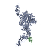
 TOXIN / microbiology / pore forming region / CROP dynamics /
  Function and homology information
 Authors
 Sweden,
  Denmark, 11items
  Citation
 Structure visualization
 Downloads & links
 Other downloads


 PDBj
PDBj


 Assembly

 Mass: 65.409 Da / Num. of mol.: 1 / Source method: obtained synthetically / Formula: Zn
 Sample preparation
 Electron microscopy imaging
 Processing