 Movie
Movie Controller

[English] 日本語
 Yorodumi
- PDB-8pyz: Structure of Ompk36GD from Klebsiella pneumonia, solved at wavele... -
+ Open data

- Basic information
| Entry | Database: PDB / ID: 8pyz | ||||||
|---|---|---|---|---|---|---|---|
| Title | Structure of Ompk36GD from Klebsiella pneumonia, solved at wavelength 4.13 A | ||||||
 Components Components | OmpK36 | ||||||
 Keywords Keywords |  MEMBRANE PROTEIN / carbapenemase MEMBRANE PROTEIN / carbapenemase | ||||||
| Function / homology |  Function and homology informationporin activity / pore complex / monoatomic ion transmembrane transport / cell outer membrane Function and homology informationporin activity / pore complex / monoatomic ion transmembrane transport / cell outer membraneSimilarity search - Function | ||||||
| Biological species |  Klebsiella pneumoniae (bacteria) Klebsiella pneumoniae (bacteria) | ||||||
| Method | X-RAY DIFFRACTION / SYNCHROTRON / SAD / Resolution: 2.7 Å | ||||||
 Authors Authors | Duman, R. / El Omari, K. / Mykhaylyk, V. / Orr, C. / Kwong, H. / Beis, K. / Wagner, A. | ||||||
| Funding support |  United Kingdom, 1items United Kingdom, 1items
| ||||||
 Citation Citation | Journal: Commun Chem / Year: 2023 Title: Experimental phasing opportunities for macromolecular crystallography at very long wavelengths. Authors: El Omari, K. / Duman, R. / Mykhaylyk, V. / Orr, C.M. / Latimer-Smith, M. / Winter, G. / Grama, V. / Qu, F. / Bountra, K. / Kwong, H.S. / Romano, M. / Reis, R.I. / Vogeley, L. / Vecchia, L. / ...Authors: El Omari, K. / Duman, R. / Mykhaylyk, V. / Orr, C.M. / Latimer-Smith, M. / Winter, G. / Grama, V. / Qu, F. / Bountra, K. / Kwong, H.S. / Romano, M. / Reis, R.I. / Vogeley, L. / Vecchia, L. / Owen, C.D. / Wittmann, S. / Renner, M. / Senda, M. / Matsugaki, N. / Kawano, Y. / Bowden, T.A. / Moraes, I. / Grimes, J.M. / Mancini, E.J. / Walsh, M.A. / Guzzo, C.R. / Owens, R.J. / Jones, E.Y. / Brown, D.G. / Stuart, D.I. / Beis, K. / Wagner, A. | ||||||
| History |
|
- Structure visualization
| Structure viewer | Molecule: MolmilJmol/JSmol |
|---|
- Downloads & links
-Download
| PDBx/mmCIF format | 8pyz.cif.gz | 489.7 KB | Display | PDBx/mmCIF format |
|---|---|---|---|---|
| PDB format | pdb8pyz.ent.gz | 336.5 KB | Display | PDB format |
| PDBx/mmJSON format | 8pyz.json.gz | Tree view | PDBx/mmJSON format | |
| Others |  Other downloads Other downloads |
-Validation report
| Arichive directory | https://data.pdbj.org/pub/pdb/validation_reports/py/8pyzftp://data.pdbj.org/pub/pdb/validation_reports/py/8pyz | HTTPS FTP |
|---|
-Related structure data
| Related structure data | 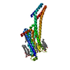 8pwnC 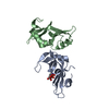 8px0C 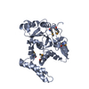 8px1C 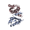 8px4C 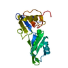 8px5C  8px7C  8px9C 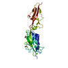 8pxcC  8pxgC  8pxhC  8pxjC  8pxkC 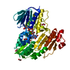 8pxlC 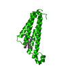 8pyvC 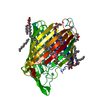 8pz4C 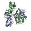 8pz5C C: citing same article ( |
|---|---|
| Similar structure data |
-Links
 PDBj
PDBj- Assembly
| Deposited unit | 
| |||||||||||||||||||||||||||||||||||||||||||||||||||||||||||||||||||||||||||||||||||||||||
|---|---|---|---|---|---|---|---|---|---|---|---|---|---|---|---|---|---|---|---|---|---|---|---|---|---|---|---|---|---|---|---|---|---|---|---|---|---|---|---|---|---|---|---|---|---|---|---|---|---|---|---|---|---|---|---|---|---|---|---|---|---|---|---|---|---|---|---|---|---|---|---|---|---|---|---|---|---|---|---|---|---|---|---|---|---|---|---|---|---|---|
| 1 |
| |||||||||||||||||||||||||||||||||||||||||||||||||||||||||||||||||||||||||||||||||||||||||
| Unit cell |
| |||||||||||||||||||||||||||||||||||||||||||||||||||||||||||||||||||||||||||||||||||||||||
| Noncrystallographic symmetry (NCS) | NCS domain:
NCS domain segments: Ens-ID: 1
|