 Movie
Movie Controller


+ Open data

- Basic information
| Entry | Database: PDB / ID: 8px0 | ||||||
|---|---|---|---|---|---|---|---|
| Title | Structure of ribonuclease A, solved at wavelength 2.75 A | ||||||
 Components Components | Ribonuclease pancreatic Pancreatic ribonuclease family Pancreatic ribonuclease family | ||||||
 Keywords Keywords | MEMBRANE PROTEIN / RNAse A | ||||||
| Function / homology |  Function and homology informationpancreatic ribonuclease / ribonuclease A activity / RNA nuclease activity / nucleic acid binding / lyase activity / defense response to Gram-positive bacterium / extracellular region Function and homology informationpancreatic ribonuclease / ribonuclease A activity / RNA nuclease activity / nucleic acid binding / lyase activity / defense response to Gram-positive bacterium / extracellular regionSimilarity search - Function | ||||||
| Biological species |  Bos taurus (cattle) Bos taurus (cattle) | ||||||
| Method | X-RAY DIFFRACTION / SYNCHROTRON / SAD / Resolution: 1.8 Å | ||||||
 Authors Authors | El Omari, K. / Duman, R. / Mykhaylyk, V. / Orr, C. / Romano, M. / Beis, K. / Wagner, A. | ||||||
| Funding support |  United Kingdom, 1items United Kingdom, 1items
| ||||||
 Citation Citation | Journal: Commun Chem / Year: 2023 Title: Experimental phasing opportunities for macromolecular crystallography at very long wavelengths. Authors: El Omari, K. / Duman, R. / Mykhaylyk, V. / Orr, C.M. / Latimer-Smith, M. / Winter, G. / Grama, V. / Qu, F. / Bountra, K. / Kwong, H.S. / Romano, M. / Reis, R.I. / Vogeley, L. / Vecchia, L. / ...Authors: El Omari, K. / Duman, R. / Mykhaylyk, V. / Orr, C.M. / Latimer-Smith, M. / Winter, G. / Grama, V. / Qu, F. / Bountra, K. / Kwong, H.S. / Romano, M. / Reis, R.I. / Vogeley, L. / Vecchia, L. / Owen, C.D. / Wittmann, S. / Renner, M. / Senda, M. / Matsugaki, N. / Kawano, Y. / Bowden, T.A. / Moraes, I. / Grimes, J.M. / Mancini, E.J. / Walsh, M.A. / Guzzo, C.R. / Owens, R.J. / Jones, E.Y. / Brown, D.G. / Stuart, D.I. / Beis, K. / Wagner, A. | ||||||
| History |
|
- Structure visualization
| Structure viewer | Molecule: MolmilJmol/JSmol |
|---|
- Downloads & links
-Download
| PDBx/mmCIF format | 8px0.cif.gz | 113.2 KB | Display | PDBx/mmCIF format |
|---|---|---|---|---|
| PDB format | pdb8px0.ent.gz | 85.9 KB | Display | PDB format |
| PDBx/mmJSON format | 8px0.json.gz | Tree view | PDBx/mmJSON format | |
| Others |  Other downloads Other downloads |
-Validation report
| Arichive directory | https://data.pdbj.org/pub/pdb/validation_reports/px/8px0ftp://data.pdbj.org/pub/pdb/validation_reports/px/8px0 | HTTPS FTP |
|---|
-Related structure data
| Related structure data | 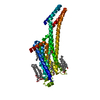 8pwnC 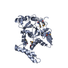 8px1C 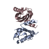 8px4C 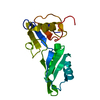 8px5C 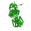 8px7C  8px9C 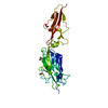 8pxcC  8pxgC 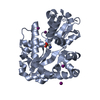 8pxhC  8pxjC  8pxkC 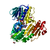 8pxlC 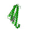 8pyvC 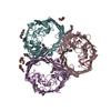 8pyzC 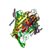 8pz4C 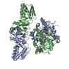 8pz5C C: citing same article ( |
|---|---|
| Similar structure data |
-Links
 PDBj
PDBj

- Assembly
| Deposited unit | 
| ||||||||
|---|---|---|---|---|---|---|---|---|---|
| 1 |
| ||||||||
| 2 |
| ||||||||
| Unit cell |
| ||||||||
| Components on special symmetry positions |
|
-Components
| #1: Protein | Pancreatic ribonuclease family / RNase 1 / RNase A Mass: 13708.326 Da / Num. of mol.: 2 Source method: isolated from a genetically manipulated source Source: (gene. exp.) Bos taurus (cattle) / Gene: RNASE1, RNS1 / Production host:  Escherichia coli (E. coli) / References: UniProt: P61823, pancreatic ribonuclease Escherichia coli (E. coli) / References: UniProt: P61823, pancreatic ribonuclease#2: Chemical | ChemComp-0U / | Uridine monophosphate  Type: L-RNA linking / Mass: 324.181 Da / Num. of mol.: 1 / Source method: isolated from a natural source / Formula: C9H13N2O9P Type: L-RNA linking / Mass: 324.181 Da / Num. of mol.: 1 / Source method: isolated from a natural source / Formula: C9H13N2O9P#3: Water | ChemComp-HOH / | Water Mass: 18.015 Da / Num. of mol.: 139 / Source method: isolated from a natural source / Formula: H2O Mass: 18.015 Da / Num. of mol.: 139 / Source method: isolated from a natural source / Formula: H2OHas ligand of interest | N | |
|---|
-Experimental details
-Experiment
| Experiment | Method: X-RAY DIFFRACTION / Number of used crystals: 1 |
|---|
- Sample preparation
| Crystal | Density Matthews: 2.16 Å3/Da / Density % sol: 43.11 % |
|---|---|
| Crystal grow | Temperature: 293 K / Method: vapor diffusion, sitting drop / Details: 20 mM sodium citrate pH 5.5, 28 % PEG 4000 |
-Data collection
| Diffraction | Mean temperature: 80 K / Serial crystal experiment: N |
|---|---|
| Diffraction source | Source: SYNCHROTRON / Site: Diamond / Beamline: I23 / Wavelength: 2.7552 Å |
| Detector | Type: DECTRIS PILATUS 12M / Detector: PIXEL / Date: Aug 1, 2019 |
| Radiation | Protocol: SINGLE WAVELENGTH / Monochromatic (M) / Laue (L): M / Scattering type: x-ray |
| Radiation wavelength | Wavelength: 2.7552 Å / Relative weight: 1 |
| Reflection | Resolution: 1.8→50.01 Å / Num. obs: 17977 / % possible obs: 81.29 % / Redundancy: 17.5 % / CC1/2: 0.995 / Rmerge(I) obs: 0.09291 / Rpim(I) all: 0.02108 / Rrim(I) all: 0.09543 / Net I/σ(I): 24.67 |
| Reflection shell | Resolution: 1.8→1.864 Å / Redundancy: 8.2 % / Rmerge(I) obs: 0.3414 / Mean I/σ(I) obs: 5.03 / Num. unique obs: 1412 / CC1/2: 0.915 / Rpim(I) all: 0.12 / Rrim(I) all: 0.3644 / % possible all: 65.04 |
- Processing
| Software |
| ||||||||||||||||||||||||||||||||||||||||||||||||||||||||||||||||||||||||||||||||||||||||||||||||||||||||||||||||||||||||||||||||||||||||||||||||||||||||||||||||||||||||||||||||||||||
|---|---|---|---|---|---|---|---|---|---|---|---|---|---|---|---|---|---|---|---|---|---|---|---|---|---|---|---|---|---|---|---|---|---|---|---|---|---|---|---|---|---|---|---|---|---|---|---|---|---|---|---|---|---|---|---|---|---|---|---|---|---|---|---|---|---|---|---|---|---|---|---|---|---|---|---|---|---|---|---|---|---|---|---|---|---|---|---|---|---|---|---|---|---|---|---|---|---|---|---|---|---|---|---|---|---|---|---|---|---|---|---|---|---|---|---|---|---|---|---|---|---|---|---|---|---|---|---|---|---|---|---|---|---|---|---|---|---|---|---|---|---|---|---|---|---|---|---|---|---|---|---|---|---|---|---|---|---|---|---|---|---|---|---|---|---|---|---|---|---|---|---|---|---|---|---|---|---|---|---|---|---|---|---|
| Refinement | Method to determine structure: SAD / Resolution: 1.8→50.01 Å / Cor.coef. Fo:Fc: 0.953 / Cor.coef. Fo:Fc free: 0.94 / SU B: 6.065 / SU ML: 0.097 / Cross valid method: THROUGHOUT / ESU R: 0.188 / ESU R Free: 0.162 / Stereochemistry target values: MAXIMUM LIKELIHOOD / Details: HYDROGENS HAVE BEEN ADDED IN THE RIDING POSITIONS
| ||||||||||||||||||||||||||||||||||||||||||||||||||||||||||||||||||||||||||||||||||||||||||||||||||||||||||||||||||||||||||||||||||||||||||||||||||||||||||||||||||||||||||||||||||||||
| Solvent computation | Ion probe radii: 0.8 Å / Shrinkage radii: 0.8 Å / VDW probe radii: 1.2 Å / Solvent model: MASK | ||||||||||||||||||||||||||||||||||||||||||||||||||||||||||||||||||||||||||||||||||||||||||||||||||||||||||||||||||||||||||||||||||||||||||||||||||||||||||||||||||||||||||||||||||||||
| Displacement parameters | Biso mean: 33.321 Å2
| ||||||||||||||||||||||||||||||||||||||||||||||||||||||||||||||||||||||||||||||||||||||||||||||||||||||||||||||||||||||||||||||||||||||||||||||||||||||||||||||||||||||||||||||||||||||
| Refinement step | Cycle: 1 / Resolution: 1.8→50.01 Å
| ||||||||||||||||||||||||||||||||||||||||||||||||||||||||||||||||||||||||||||||||||||||||||||||||||||||||||||||||||||||||||||||||||||||||||||||||||||||||||||||||||||||||||||||||||||||
| Refine LS restraints |
|