 Movie
Movie Controller


+ Open data

- Basic information
| Entry | Database: PDB / ID: 8pqh | |||||||||||||||
|---|---|---|---|---|---|---|---|---|---|---|---|---|---|---|---|---|
| Title | PDGFRA T674I mutant kinase domain in complex with avapritinib | |||||||||||||||
 Components Components | Platelet-derived growth factor receptor alpha PDGFRA PDGFRA | |||||||||||||||
 Keywords Keywords | TRANSFERASE / PDGFRA protein kinase / avapritinib / inhibitor / tyrosine kinase / transmembrane receptor / platelet-derived growth factor receptor alpha / PDGF / platelet-derived growth factor / GIST / gastrointestinal stromal tumors / gatekeeper mutant / P16234 | |||||||||||||||
| Function / homology |  Function and homology information Function and homology informationplatelet-derived growth factor alpha-receptor activity / platelet-derived growth factor receptor-alpha signaling pathway / Imatinib-resistant PDGFR mutants / Sunitinib-resistant PDGFR mutants / Regorafenib-resistant PDGFR mutants / Sorafenib-resistant PDGFR mutants / PDGFR mutants bind TKIs / metanephric glomerular capillary formation / regulation of mesenchymal stem cell differentiation / luteinization ...platelet-derived growth factor alpha-receptor activity / platelet-derived growth factor receptor-alpha signaling pathway / Imatinib-resistant PDGFR mutants / Sunitinib-resistant PDGFR mutants / Regorafenib-resistant PDGFR mutants / Sorafenib-resistant PDGFR mutants / PDGFR mutants bind TKIs / metanephric glomerular capillary formation / regulation of mesenchymal stem cell differentiation / luteinization / positive regulation of cell proliferation by VEGF-activated platelet derived growth factor receptor signaling pathway / platelet-derived growth factor binding / embryonic skeletal system morphogenesis / vascular endothelial growth factor binding / retina vasculature development in camera-type eye / cardiac myofibril assembly / embryonic digestive tract morphogenesis / vascular endothelial growth factor receptor activity / Leydig cell differentiation / cell activation / male genitalia development / positive regulation of chemotaxis / Signaling by PDGF / signal transduction involved in regulation of gene expression / embryonic cranial skeleton morphogenesis / platelet-derived growth factor receptor binding / face morphogenesis / estrogen metabolic process / adrenal gland development / roof of mouth development / odontogenesis of dentin-containing tooth / microvillus / platelet-derived growth factor receptor signaling pathway / white fat cell differentiation / negative regulation of platelet activation / hematopoietic progenitor cell differentiation / : / positive regulation of calcium-mediated signaling / Signaling by PDGFRA transmembrane, juxtamembrane and kinase domain mutants / Signaling by PDGFRA extracellular domain mutants / transmembrane receptor protein tyrosine kinase activity / extracellular matrix organization / cell chemotaxis / Downstream signal transduction / regulation of actin cytoskeleton organization / cellular response to amino acid stimulus / lung development / wound healing / cilium / receptor protein-tyrosine kinase / platelet aggregation / cellular response to reactive oxygen species / peptidyl-tyrosine phosphorylation / Constitutive Signaling by Aberrant PI3K in Cancer / positive regulation of fibroblast proliferation / PIP3 activates AKT signaling / cell junction / PI5P, PP2A and IER3 Regulate PI3K/AKT Signaling / RAF/MAP kinase cascade / in utero embryonic development / protein autophosphorylation / positive regulation of phosphatidylinositol 3-kinase/protein kinase B signal transduction / positive regulation of ERK1 and ERK2 cascade / receptor complex / protein kinase activity / positive regulation of cell migration / external side of plasma membrane / positive regulation of cell population proliferation / protein-containing complex binding / endoplasmic reticulum membrane / Golgi apparatus / protein homodimerization activity / protein-containing complex / nucleoplasm / ATP binding / membrane / nucleus / plasma membrane / cytoplasmSimilarity search - Function | |||||||||||||||
| Biological species |  Homo sapiens (human) Homo sapiens (human) | |||||||||||||||
| Method | X-RAY DIFFRACTION / SYNCHROTRON / MOLECULAR REPLACEMENT / Resolution: 2.5 Å | |||||||||||||||
 Authors Authors | Teuber, A. / Kleinboelting, S. / Mueller, M.P. / Rauh, D. | |||||||||||||||
| Funding support | European Union,  Germany, 4items Germany, 4items
| |||||||||||||||
 Citation Citation | Journal: Nat Commun / Year: 2024 Title: Avapritinib-based SAR studies unveil a binding pocket in KIT and PDGFRA. Authors: Teuber, A. / Schulz, T. / Fletcher, B.S. / Gontla, R. / Muhlenberg, T. / Zischinsky, M.L. / Niggenaber, J. / Weisner, J. / Kleinbolting, S.B. / Lategahn, J. / Sievers, S. / Muller, M.P. / Bauer, S. / Rauh, D. | |||||||||||||||
| History |
|
- Structure visualization
| Structure viewer | Molecule: MolmilJmol/JSmol |
|---|
- Downloads & links
-Download
| PDBx/mmCIF format | 8pqh.cif.gz | 167.2 KB | Display | PDBx/mmCIF format |
|---|---|---|---|---|
| PDB format | pdb8pqh.ent.gz | 108.3 KB | Display | PDB format |
| PDBx/mmJSON format | 8pqh.json.gz | Tree view | PDBx/mmJSON format | |
| Others |  Other downloads Other downloads |
-Validation report
| Arichive directory | https://data.pdbj.org/pub/pdb/validation_reports/pq/8pqhftp://data.pdbj.org/pub/pdb/validation_reports/pq/8pqh | HTTPS FTP |
|---|
-Related structure data
| Related structure data | 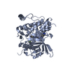 8pq9C 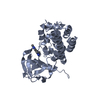 8pqaC  8pqbC 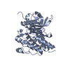 8pqcC  8pqdC  8pqeC 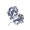 8pqfC  8pqgC 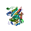 8pqiC 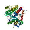 8pqjC 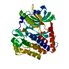 8pqkC C: citing same article ( |
|---|---|
| Similar structure data | |
| Experimental dataset #1 | Data set type: diffraction image data / Details: https://www.proteindiffraction.org/project/8PQH/ |
-Links
 PDBj
PDBj


- Assembly
| Deposited unit | 
| ||||||||||||
|---|---|---|---|---|---|---|---|---|---|---|---|---|---|
| 1 |
| ||||||||||||
| Unit cell |
|
-Components
| #1: Protein | PDGFRA / PDGF-R-alpha / PDGFR-alpha / Alpha platelet-derived growth factor receptor / Alpha-type platelet- ...PDGF-R-alpha / PDGFR-alpha / Alpha platelet-derived growth factor receptor / Alpha-type platelet-derived growth factor receptor / CD140 antigen-like family member A / CD140a antigen / Platelet-derived growth factor alpha receptor / Platelet-derived growth factor receptor 2 / PDGFR-2 Mass: 40226.371 Da / Num. of mol.: 1 / Mutation: T674I Source method: isolated from a genetically manipulated source Source: (gene. exp.) Homo sapiens (human) / Gene: PDGFRA, PDGFR2, RHEPDGFRA / Production host:   Spodoptera frugiperda (fall armyworm) Spodoptera frugiperda (fall armyworm)References: UniProt: P16234, receptor protein-tyrosine kinase |
|---|---|
| #2: Chemical | ChemComp-9JI / (  Mass: 498.558 Da / Num. of mol.: 1 / Source method: obtained synthetically / Formula: C26H27FN10 / Feature type: SUBJECT OF INVESTIGATION Mass: 498.558 Da / Num. of mol.: 1 / Source method: obtained synthetically / Formula: C26H27FN10 / Feature type: SUBJECT OF INVESTIGATION |
| #3: Chemical | ChemComp-PEG / Diethylene glycol  Mass: 106.120 Da / Num. of mol.: 1 / Source method: obtained synthetically / Formula: C4H10O3 Mass: 106.120 Da / Num. of mol.: 1 / Source method: obtained synthetically / Formula: C4H10O3 |
| #4: Water | ChemComp-HOH / Water Mass: 18.015 Da / Num. of mol.: 22 / Source method: isolated from a natural source / Formula: H2O Mass: 18.015 Da / Num. of mol.: 22 / Source method: isolated from a natural source / Formula: H2O |
| Has ligand of interest | Y |
-Experimental details
-Experiment
| Experiment | Method: X-RAY DIFFRACTION / Number of used crystals: 1 |
|---|
- Sample preparation
| Crystal | Density Matthews: 2.02 Å3/Da / Density % sol: 39.24 % |
|---|---|
| Crystal grow | Temperature: 293.15 K / Method: vapor diffusion, hanging drop / Details: 10 mg/mL, 13% PEG3350, 200 mM KCl |
-Data collection
| Diffraction | Mean temperature: 100 K / Serial crystal experiment: N |
|---|---|
| Diffraction source | Source: SYNCHROTRON / Site: SLS  / Beamline: X10SA / Wavelength: 1 Å / Beamline: X10SA / Wavelength: 1 Å |
| Detector | Type: DECTRIS EIGER2 X 16M / Detector: PIXEL / Date: Feb 17, 2021 |
| Radiation | Monochromator: Si(111) / Protocol: SINGLE WAVELENGTH / Monochromatic (M) / Laue (L): M / Scattering type: x-ray |
| Radiation wavelength | Wavelength: 1 Å / Relative weight: 1 |
| Reflection | Resolution: 2.5→46.75 Å / Num. obs: 14185 / % possible obs: 100 % / Redundancy: 13.09 % / Biso Wilson estimate: 71.45 Å2 / CC1/2: 0.999 / Rrim(I) all: 0.08 / Net I/σ(I): 18.79 |
| Reflection shell | Resolution: 2.5→2.6 Å / Redundancy: 13.67 % / Mean I/σ(I) obs: 2.06 / Num. unique obs: 1529 / CC1/2: 0.77 / Rrim(I) all: 1.499 / % possible all: 100 |
- Processing
| Software |
| ||||||||||||||||||||||||||||||||||||||||||||||||||||||||||||||||||||||||||||||||||||||||||||||||||||
|---|---|---|---|---|---|---|---|---|---|---|---|---|---|---|---|---|---|---|---|---|---|---|---|---|---|---|---|---|---|---|---|---|---|---|---|---|---|---|---|---|---|---|---|---|---|---|---|---|---|---|---|---|---|---|---|---|---|---|---|---|---|---|---|---|---|---|---|---|---|---|---|---|---|---|---|---|---|---|---|---|---|---|---|---|---|---|---|---|---|---|---|---|---|---|---|---|---|---|---|---|---|
| Refinement | Method to determine structure: MOLECULAR REPLACEMENT / Resolution: 2.5→46.57 Å / SU ML: 0.399 / Cross valid method: FREE R-VALUE / σ(F): 1.36 / Phase error: 30.9463 Stereochemistry target values: GeoStd + Monomer Library + CDL v1.2
| ||||||||||||||||||||||||||||||||||||||||||||||||||||||||||||||||||||||||||||||||||||||||||||||||||||
| Solvent computation | Shrinkage radii: 0.9 Å / VDW probe radii: 1.11 Å / Solvent model: FLAT BULK SOLVENT MODEL | ||||||||||||||||||||||||||||||||||||||||||||||||||||||||||||||||||||||||||||||||||||||||||||||||||||
| Displacement parameters | Biso mean: 78.98 Å2 | ||||||||||||||||||||||||||||||||||||||||||||||||||||||||||||||||||||||||||||||||||||||||||||||||||||
| Refinement step | Cycle: LAST / Resolution: 2.5→46.57 Å
| ||||||||||||||||||||||||||||||||||||||||||||||||||||||||||||||||||||||||||||||||||||||||||||||||||||
| Refine LS restraints |
| ||||||||||||||||||||||||||||||||||||||||||||||||||||||||||||||||||||||||||||||||||||||||||||||||||||
| LS refinement shell |
| ||||||||||||||||||||||||||||||||||||||||||||||||||||||||||||||||||||||||||||||||||||||||||||||||||||
| Refinement TLS params. | Method: refined / Refine-ID: X-RAY DIFFRACTION
| ||||||||||||||||||||||||||||||||||||||||||||||||||||||||||||||||||||||||||||||||||||||||||||||||||||
| Refinement TLS group | Refine-ID: X-RAY DIFFRACTION / Auth asym-ID: A / Label asym-ID: A
|
