 Movie
Movie Controller

[English] 日本語
 Yorodumi
- PDB-8oy9: Time-resolved SFX structure of the class II photolyase complexed ... -
+ Open data

- Basic information
| Entry | Database: PDB / ID: 8oy9 | |||||||||
|---|---|---|---|---|---|---|---|---|---|---|
| Title | Time-resolved SFX structure of the class II photolyase complexed with a thymine dimer (1 microsecond pump-probe delay) | |||||||||
 Components Components |
| |||||||||
 Keywords Keywords |  LYASE / DNA binding protein / DNA repair enzyme / flavoprotein / photoenzyme LYASE / DNA binding protein / DNA repair enzyme / flavoprotein / photoenzyme | |||||||||
| Function / homology |  Function and homology informationdeoxyribodipyrimidine photo-lyase / deoxyribodipyrimidine photo-lyase activity / nucleotide binding / DNA repair / DNA binding Function and homology informationdeoxyribodipyrimidine photo-lyase / deoxyribodipyrimidine photo-lyase activity / nucleotide binding / DNA repair / DNA bindingSimilarity search - Function | |||||||||
| Biological species |  Methanosarcina mazei Go1 (archaea) Methanosarcina mazei Go1 (archaea)synthetic construct (others) | |||||||||
| Method | X-RAY DIFFRACTION / FREE ELECTRON LASER / MOLECULAR REPLACEMENT / Resolution: 2.24 Å | |||||||||
 Authors Authors | Lane, T.J. / Christou, N.-E. / Melo, D.V.M. / Apostolopoulou, V. / Pateras, A. / Mashhour, A.R. / Galchenkova, M. / Gunther, S. / Reinke, P. / Kremling, V. ...Lane, T.J. / Christou, N.-E. / Melo, D.V.M. / Apostolopoulou, V. / Pateras, A. / Mashhour, A.R. / Galchenkova, M. / Gunther, S. / Reinke, P. / Kremling, V. / Oberthuer, D. / Henkel, A. / Sprenger, J. / Scheer, T.E.S. / Lange, E. / Yefanov, O.N. / Middendorf, P. / Sellberg, J.A. / Schubert, R. / Fadini, A. / Cirelli, C. / Beale, E.V. / Johnson, P. / Dworkowski, F. / Ozerov, D. / Bertrand, Q. / Wranik, M. / Zitter, E.D. / Turk, D. / Bajt, S. / Chapman, H. / Bacellar, C. | |||||||||
| Funding support |  Germany, Germany,  Switzerland, 2items Switzerland, 2items
| |||||||||
 Citation Citation | Journal: Science / Year: 2023 Title: Time-resolved crystallography captures light-driven DNA repair. Authors: Christou, N.E. / Apostolopoulou, V. / Melo, D.V.M. / Ruppert, M. / Fadini, A. / Henkel, A. / Sprenger, J. / Oberthuer, D. / Gunther, S. / Pateras, A. / Rahmani Mashhour, A. / Yefanov, O.M. / ...Authors: Christou, N.E. / Apostolopoulou, V. / Melo, D.V.M. / Ruppert, M. / Fadini, A. / Henkel, A. / Sprenger, J. / Oberthuer, D. / Gunther, S. / Pateras, A. / Rahmani Mashhour, A. / Yefanov, O.M. / Galchenkova, M. / Reinke, P.Y.A. / Kremling, V. / Scheer, T.E.S. / Lange, E.R. / Middendorf, P. / Schubert, R. / De Zitter, E. / Lumbao-Conradson, K. / Herrmann, J. / Rahighi, S. / Kunavar, A. / Beale, E.V. / Beale, J.H. / Cirelli, C. / Johnson, P.J.M. / Dworkowski, F. / Ozerov, D. / Bertrand, Q. / Wranik, M. / Bacellar, C. / Bajt, S. / Wakatsuki, S. / Sellberg, J.A. / Huse, N. / Turk, D. / Chapman, H.N. / Lane, T.J. | |||||||||
| History |
|
- Structure visualization
| Structure viewer | Molecule: MolmilJmol/JSmol |
|---|
- Downloads & links
-Download
| PDBx/mmCIF format | 8oy9.cif.gz | 523.5 KB | Display | PDBx/mmCIF format |
|---|---|---|---|---|
| PDB format | pdb8oy9.ent.gz | 354.8 KB | Display | PDB format |
| PDBx/mmJSON format | 8oy9.json.gz | Tree view | PDBx/mmJSON format | |
| Others |  Other downloads Other downloads |
-Validation report
| Arichive directory | https://data.pdbj.org/pub/pdb/validation_reports/oy/8oy9ftp://data.pdbj.org/pub/pdb/validation_reports/oy/8oy9 | HTTPS FTP |
|---|
-Related structure data
| Related structure data | 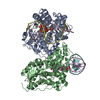 8oetC  8oy3C 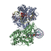 8oy4C  8oy5C 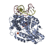 8oy6C 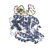 8oy7C 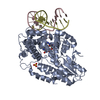 8oy8C 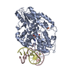 8oyaC 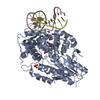 8oybC 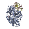 8oycC C: citing same article ( |
|---|---|
| Similar structure data | |
| Experimental dataset #1 | Data reference: 10.16907/87a9f7a4-073e-430c-aa7f-6a194897074f Data set type: diffraction image data |
-Links
 PDBj
PDBj


- Assembly
| Deposited unit | 
| ||||||||||||
|---|---|---|---|---|---|---|---|---|---|---|---|---|---|
| 1 | 
| ||||||||||||
| 2 | 
| ||||||||||||
| Unit cell |
|
-Components
-Protein , 1 types, 2 molecules AB
| #1: Protein | Photolyase / DNA photolyase Mass: 57039.734 Da / Num. of mol.: 2 Source method: isolated from a genetically manipulated source Source: (gene. exp.) Methanosarcina mazei Go1 (archaea) / Gene: MM_0852 / Production host:  Escherichia coli BL21(DE3) (bacteria) Escherichia coli BL21(DE3) (bacteria)References: UniProt: Q8PYK9, deoxyribodipyrimidine photo-lyase |
|---|
-DNA chain , 2 types, 4 molecules CEDF
| #2: DNA chain | Mass: 4256.767 Da / Num. of mol.: 2 / Source method: obtained synthetically / Source: (synth.) synthetic construct (others) #3: DNA chain | Mass: 4305.805 Da / Num. of mol.: 2 / Source method: obtained synthetically / Source: (synth.) synthetic construct (others) |
|---|
-Non-polymers , 3 types, 181 molecules 


| #4: Chemical | ChemComp-SO4 / Sulfate Mass: 96.063 Da / Num. of mol.: 1 / Source method: obtained synthetically / Formula: SO4 Mass: 96.063 Da / Num. of mol.: 1 / Source method: obtained synthetically / Formula: SO4 | ||
|---|---|---|---|
| #5: Chemical |  Mass: 787.566 Da / Num. of mol.: 2 / Source method: obtained synthetically / Formula: C27H35N9O15P2 Mass: 787.566 Da / Num. of mol.: 2 / Source method: obtained synthetically / Formula: C27H35N9O15P2#6: Water | ChemComp-HOH / | WaterMass: 18.015 Da / Num. of mol.: 178 / Source method: isolated from a natural source / Formula: H2O |
-Details
| Has ligand of interest | N |
|---|
-Experimental details
-Experiment
| Experiment | Method: X-RAY DIFFRACTION / Number of used crystals: 1 |
|---|
- Sample preparation
| Crystal | Density Matthews: 2.69 Å3/Da / Density % sol: 54.19 % |
|---|---|
| Crystal grow | Temperature: 293 K / Method: batch mode Details: 100 mM Amm sulfate, 100 mM Acetate pH 4.6, 4% PEG 4000, 10 mM Tris pH 7.5, 75 mM NaCl |
-Data collection
| Diffraction | Mean temperature: 293 K / Serial crystal experiment: Y | ||||||||||||||||||||||||||||||||||||||||||||||||||||||||||||||||||||||||||||||||||||||||||||||||||||||||||||||||||||||||||||||||||||||||||||||||||||||||||||||||
|---|---|---|---|---|---|---|---|---|---|---|---|---|---|---|---|---|---|---|---|---|---|---|---|---|---|---|---|---|---|---|---|---|---|---|---|---|---|---|---|---|---|---|---|---|---|---|---|---|---|---|---|---|---|---|---|---|---|---|---|---|---|---|---|---|---|---|---|---|---|---|---|---|---|---|---|---|---|---|---|---|---|---|---|---|---|---|---|---|---|---|---|---|---|---|---|---|---|---|---|---|---|---|---|---|---|---|---|---|---|---|---|---|---|---|---|---|---|---|---|---|---|---|---|---|---|---|---|---|---|---|---|---|---|---|---|---|---|---|---|---|---|---|---|---|---|---|---|---|---|---|---|---|---|---|---|---|---|---|---|---|---|
| Diffraction source | Source: FREE ELECTRON LASER / Site: SwissFEL ARAMIS / Beamline: ESA / Wavelength: 1.035 Å | ||||||||||||||||||||||||||||||||||||||||||||||||||||||||||||||||||||||||||||||||||||||||||||||||||||||||||||||||||||||||||||||||||||||||||||||||||||||||||||||||
| Detector | Type: PSI JUNGFRAU 16M / Detector: PIXEL / Date: Jul 2, 2022 | ||||||||||||||||||||||||||||||||||||||||||||||||||||||||||||||||||||||||||||||||||||||||||||||||||||||||||||||||||||||||||||||||||||||||||||||||||||||||||||||||
| Radiation | Protocol: SINGLE WAVELENGTH / Monochromatic (M) / Laue (L): M / Scattering type: x-ray | ||||||||||||||||||||||||||||||||||||||||||||||||||||||||||||||||||||||||||||||||||||||||||||||||||||||||||||||||||||||||||||||||||||||||||||||||||||||||||||||||
| Radiation wavelength | Wavelength: 1.035 Å / Relative weight: 1 | ||||||||||||||||||||||||||||||||||||||||||||||||||||||||||||||||||||||||||||||||||||||||||||||||||||||||||||||||||||||||||||||||||||||||||||||||||||||||||||||||
| Reflection | Resolution: 2.24→31.29 Å / Num. obs: 68821 / % possible obs: 100 % / Redundancy: 920.008646 % / Biso Wilson estimate: 36.07 Å2 / CC1/2: 0.9889675 / CC star: 0.9972227 / R split: 0.0901 / Net I/σ(I): 9.496358 / Num. measured all: 63315915 | ||||||||||||||||||||||||||||||||||||||||||||||||||||||||||||||||||||||||||||||||||||||||||||||||||||||||||||||||||||||||||||||||||||||||||||||||||||||||||||||||
| Reflection shell | % possible all: 100
| ||||||||||||||||||||||||||||||||||||||||||||||||||||||||||||||||||||||||||||||||||||||||||||||||||||||||||||||||||||||||||||||||||||||||||||||||||||||||||||||||
| Serial crystallography sample delivery | Method: injection |
- Processing
| Software |
| |||||||||||||||||||||||||||||||||||||||||||||||||||||||||||||||||||||||||||||||||||||||||||||||||||||||||||||||||||||||||||||||||||||||||||||||||||
|---|---|---|---|---|---|---|---|---|---|---|---|---|---|---|---|---|---|---|---|---|---|---|---|---|---|---|---|---|---|---|---|---|---|---|---|---|---|---|---|---|---|---|---|---|---|---|---|---|---|---|---|---|---|---|---|---|---|---|---|---|---|---|---|---|---|---|---|---|---|---|---|---|---|---|---|---|---|---|---|---|---|---|---|---|---|---|---|---|---|---|---|---|---|---|---|---|---|---|---|---|---|---|---|---|---|---|---|---|---|---|---|---|---|---|---|---|---|---|---|---|---|---|---|---|---|---|---|---|---|---|---|---|---|---|---|---|---|---|---|---|---|---|---|---|---|---|---|---|
| Refinement | Method to determine structure: MOLECULAR REPLACEMENT / Resolution: 2.24→31.29 Å / SU ML: 0.5619 / Cross valid method: FREE R-VALUE / σ(F): 1.37 / Phase error: 48.0926 Stereochemistry target values: GeoStd + Monomer Library + CDL v1.2
| |||||||||||||||||||||||||||||||||||||||||||||||||||||||||||||||||||||||||||||||||||||||||||||||||||||||||||||||||||||||||||||||||||||||||||||||||||
| Solvent computation | Shrinkage radii: 0.9 Å / VDW probe radii: 1.1 Å / Solvent model: FLAT BULK SOLVENT MODEL | |||||||||||||||||||||||||||||||||||||||||||||||||||||||||||||||||||||||||||||||||||||||||||||||||||||||||||||||||||||||||||||||||||||||||||||||||||
| Displacement parameters | Biso mean: 53.67 Å2 | |||||||||||||||||||||||||||||||||||||||||||||||||||||||||||||||||||||||||||||||||||||||||||||||||||||||||||||||||||||||||||||||||||||||||||||||||||
| Refinement step | Cycle: LAST / Resolution: 2.24→31.29 Å
| |||||||||||||||||||||||||||||||||||||||||||||||||||||||||||||||||||||||||||||||||||||||||||||||||||||||||||||||||||||||||||||||||||||||||||||||||||
| Refine LS restraints |
| |||||||||||||||||||||||||||||||||||||||||||||||||||||||||||||||||||||||||||||||||||||||||||||||||||||||||||||||||||||||||||||||||||||||||||||||||||
| LS refinement shell |
| |||||||||||||||||||||||||||||||||||||||||||||||||||||||||||||||||||||||||||||||||||||||||||||||||||||||||||||||||||||||||||||||||||||||||||||||||||
| Refinement TLS params. | Method: refined / Refine-ID: X-RAY DIFFRACTION
| |||||||||||||||||||||||||||||||||||||||||||||||||||||||||||||||||||||||||||||||||||||||||||||||||||||||||||||||||||||||||||||||||||||||||||||||||||
| Refinement TLS group | Refine-ID: X-RAY DIFFRACTION / Label seq-ID: 1
|
