 Movie
Movie Controller


+ Open data

- Basic information
| Entry | Database: PDB / ID: 8owd | ||||||||||||
|---|---|---|---|---|---|---|---|---|---|---|---|---|---|
| Title | Lipidic amyloid-beta(1-40) fibril - polymorph L3 | ||||||||||||
 Components Components | Amyloid-beta A4 protein | ||||||||||||
 Keywords Keywords | PROTEIN FIBRIL /  amyloid-beta / fibril / lipids amyloid-beta / fibril / lipids | ||||||||||||
| Function / homology |  Function and homology information Function and homology informationGolgi-associated vesicle / clathrin-coated pit / heparin binding / growth cone / perikaryon / early endosome / Golgi apparatus / cell surface / endoplasmic reticulum / extracellular region / nucleusSimilarity search - Function | ||||||||||||
| Biological species |  Homo sapiens (human) Homo sapiens (human) | ||||||||||||
| Method | ELECTRON MICROSCOPY / helical reconstruction / cryo EM / Resolution: 3.28 Å | ||||||||||||
 Authors Authors | Frieg, B. / Han, M. / Giller, K. / Dienemann, C. / Riedel, D. / Becker, S. / Andreas, L.B. / Griesinger, C. / Schroeder, G.F. | ||||||||||||
| Funding support |  Germany, 3items Germany, 3items
| ||||||||||||
 Citation Citation | Journal: Nat Commun / Year: 2024 Title: Cryo-EM structures of lipidic fibrils of amyloid-β (1-40). Authors: Benedikt Frieg / Mookyoung Han / Karin Giller / Christian Dienemann / Dietmar Riedel / Stefan Becker / Loren B Andreas / Christian Griesinger / Gunnar F Schröder / Abstract: Alzheimer's disease (AD) is a progressive and incurable neurodegenerative disease characterized by the extracellular deposition of amyloid plaques. Investigation into the composition of these plaques ...Alzheimer's disease (AD) is a progressive and incurable neurodegenerative disease characterized by the extracellular deposition of amyloid plaques. Investigation into the composition of these plaques revealed a high amount of amyloid-β (Aβ) fibrils and a high concentration of lipids, suggesting that fibril-lipid interactions may also be relevant for the pathogenesis of AD. Therefore, we grew Aβ40 fibrils in the presence of lipid vesicles and determined their structure by cryo-electron microscopy (cryo-EM) to high resolution. The fold of the major polymorph is similar to the structure of brain-seeded fibrils reported previously. The majority of the lipids are bound to the fibrils, as we show by cryo-EM and NMR spectroscopy. This apparent lipid extraction from vesicles observed here in vitro provides structural insights into potentially disease-relevant fibril-lipid interactions. | ||||||||||||
| History |
|
- Structure visualization
| Structure viewer | Molecule: MolmilJmol/JSmol |
|---|
- Downloads & links
-Download
| PDBx/mmCIF format | 8owd.cif.gz | 45.2 KB | Display | PDBx/mmCIF format |
|---|---|---|---|---|
| PDB format | pdb8owd.ent.gz | 30.5 KB | Display | PDB format |
| PDBx/mmJSON format | 8owd.json.gz | Tree view | PDBx/mmJSON format | |
| Others |  Other downloads Other downloads |
-Validation report
| Arichive directory | https://data.pdbj.org/pub/pdb/validation_reports/ow/8owdftp://data.pdbj.org/pub/pdb/validation_reports/ow/8owd | HTTPS FTP |
|---|
-Related structure data
| Related structure data |  17234MC  8ovkC  8ovmC  8oweC  8owjC 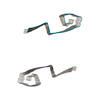 8owkC C: citing same article ( M: map data used to model this data |
|---|---|
| Similar structure data |


-Links
 PDBj
PDBj

- Assembly
| Deposited unit | 
| ||||||||||||||||||||||||||||||||||||||||||||||||||||||||
|---|---|---|---|---|---|---|---|---|---|---|---|---|---|---|---|---|---|---|---|---|---|---|---|---|---|---|---|---|---|---|---|---|---|---|---|---|---|---|---|---|---|---|---|---|---|---|---|---|---|---|---|---|---|---|---|---|---|
| 1 |
| ||||||||||||||||||||||||||||||||||||||||||||||||||||||||
| Noncrystallographic symmetry (NCS) | NCS domain:
NCS domain segments: Component-ID: 1 / Ens-ID: ens_1 / Beg auth comp-ID: ASP / Beg label comp-ID: ASP / End auth comp-ID: VAL / End label comp-ID: VAL / Auth seq-ID: 1 - 40 / Label seq-ID: 1 - 40
NCS oper:
|
-Components
| #1: Protein/peptide | Mass: 4335.852 Da / Num. of mol.: 5 Source method: isolated from a genetically manipulated source Source: (gene. exp.) Homo sapiens (human) / Production host:  Escherichia coli BL21(DE3) (bacteria) / References: UniProt: B4DM00 Escherichia coli BL21(DE3) (bacteria) / References: UniProt: B4DM00 |
|---|
-Experimental details
-Experiment
| Experiment | Method: ELECTRON MICROSCOPY |
|---|---|
| EM experiment | Aggregation state: FILAMENT / 3D reconstruction method: helical reconstruction |
- Sample preparation
| Component | Name: The L3 amyloid-beta(1-40) fibril in complex with lipids Type: COMPLEX / Entity ID: all / Source: RECOMBINANT |
|---|---|
| Molecular weight | Experimental value: NO |
| Source (natural) | Organism: Homo sapiens (human) |
| Source (recombinant) | Organism: Escherichia coli BL21(DE3) (bacteria) |
| Buffer solution | pH: 6.5 |
| Specimen | Embedding applied: NO / Shadowing applied: NO / Staining applied: NO / Vitrification applied: YES |
| Vitrification | Cryogen name: ETHANE |
- Electron microscopy imaging
| Experimental equipment |  Model: Titan Krios / Image courtesy: FEI Company |
|---|---|
| Microscopy | Model: FEI TITAN KRIOS |
| Electron gun | Electron source: FIELD EMISSION GUN / Accelerating voltage: 300 kV / Illumination mode: FLOOD BEAM |
| Electron lens | Mode: BRIGHT FIELDBright-field microscopy / Nominal defocus max: 2500 nm / Nominal defocus min: 500 nm |
| Image recording | Electron dose: 40 e/Å2 / Film or detector model: GATAN K3 (6k x 4k) |
- Processing
| Software |
| ||||||||||||||||||||||||||||||
|---|---|---|---|---|---|---|---|---|---|---|---|---|---|---|---|---|---|---|---|---|---|---|---|---|---|---|---|---|---|---|---|
| CTF correction | Type: NONE | ||||||||||||||||||||||||||||||
| Helical symmerty | Angular rotation/subunit: -2.07 ° / Axial rise/subunit: 4.67 Å / Axial symmetry: C1 | ||||||||||||||||||||||||||||||
| 3D reconstruction | Resolution: 3.28 Å / Resolution method: FSC 0.143 CUT-OFF / Num. of particles: 51100 / Symmetry type: HELICAL | ||||||||||||||||||||||||||||||
| Refinement | Cross valid method: NONE Stereochemistry target values: GeoStd + Monomer Library + CDL v1.2 | ||||||||||||||||||||||||||||||
| Displacement parameters | Biso mean: 82.97 Å2 | ||||||||||||||||||||||||||||||
| Refine LS restraints |
| ||||||||||||||||||||||||||||||
| Refine LS restraints NCS |
|
