Movie
Movie Controller

[English] 日本語
 Yorodumi
- PDB-8oud: Structure of the human neutral amino acid transporter ASCT2 in co... -
+ Open data

- Basic information
| Entry | Database: PDB / ID: 8oud | ||||||
|---|---|---|---|---|---|---|---|
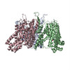
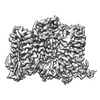
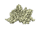
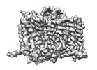
| Title | Structure of the human neutral amino acid transporter ASCT2 in complex with nanobody 469 | ||||||
 Components Components |
| ||||||
 Keywords Keywords |  MEMBRANE PROTEIN / Neutral aminoacid transmembrane transporter activity / ASCT2 / Complex / Nanobody MEMBRANE PROTEIN / Neutral aminoacid transmembrane transporter activity / ASCT2 / Complex / Nanobody | ||||||
| Function / homology |  Function and homology information Function and homology informationglutamine secretion / L-glutamine import across plasma membrane / glutamine transport / L-glutamine transmembrane transporter activity / L-serine transmembrane transporter activity / ligand-gated channel activity / neutral amino acid transport / amino acid transmembrane transporter activity / Amino acid transport across the plasma membrane / neutral L-amino acid transmembrane transporter activity ...glutamine secretion / L-glutamine import across plasma membrane / glutamine transport / L-glutamine transmembrane transporter activity / L-serine transmembrane transporter activity / ligand-gated channel activity / neutral amino acid transport / amino acid transmembrane transporter activity / Amino acid transport across the plasma membrane / neutral L-amino acid transmembrane transporter activity / L-aspartate transmembrane transporter activity / L-aspartate import across plasma membrane / symporter activity / amino acid transport / antiporter activity / RHOJ GTPase cycle / RHOQ GTPase cycle / protein homotrimerization / RHOH GTPase cycle / transport across blood-brain barrier / RAC3 GTPase cycle / RAC1 GTPase cycle / erythrocyte differentiation / basal plasma membrane / melanosome / virus receptor activity / signaling receptor activity / extracellular exosome / membrane / metal ion binding / plasma membraneSimilarity search - Function | ||||||
| Biological species |  Homo sapiens (human) Homo sapiens (human) Lama glama (llama) Lama glama (llama) | ||||||
| Method | ELECTRON MICROSCOPY / single particle reconstruction / cryo EM / Resolution: 2.31 Å | ||||||
 Authors Authors | Canul-Tec, J. / Reyes, N. | ||||||
| Funding support | European Union, 1items
| ||||||
 Citation Citation | Journal: Nat Struct Mol Biol / Year: 2024 Title: Receptor-recognition and antiviral mechanisms of retrovirus-derived human proteins. Authors: Shashank Khare / Miryam I Villalba / Juan C Canul-Tec / Arantza Balsebre Cajiao / Anand Kumar / Marija Backovic / Felix A Rey / Els Pardon / Jan Steyaert / Camilo Perez / Nicolas Reyes /     Abstract: Human syncytin-1 and suppressyn are cellular proteins of retroviral origin involved in cell-cell fusion events to establish the maternal-fetal interface in the placenta. In cell culture, they ...Human syncytin-1 and suppressyn are cellular proteins of retroviral origin involved in cell-cell fusion events to establish the maternal-fetal interface in the placenta. In cell culture, they restrict infections from members of the largest interference group of vertebrate retroviruses, and are regarded as host immunity factors expressed during development. At the core of the syncytin-1 and suppressyn functions are poorly understood mechanisms to recognize a common cellular receptor, the membrane transporter ASCT2. Here, we present cryo-electron microscopy structures of human ASCT2 in complexes with the receptor-binding domains of syncytin-1 and suppressyn. Despite their evolutionary divergence, the two placental proteins occupy similar positions in ASCT2, and are stabilized by the formation of a hybrid β-sheet or 'clamp' with the receptor. Structural predictions of the receptor-binding domains of extant retroviruses indicate overlapping binding interfaces and clamping sites with ASCT2, revealing a competition mechanism between the placental proteins and the retroviruses. Our work uncovers a common ASCT2 recognition mechanism by a large group of endogenous and disease-causing retroviruses, and provides high-resolution views on how placental human proteins exert morphological and immunological functions. | ||||||
| History |
|
- Structure visualization
| Structure viewer | Molecule: MolmilJmol/JSmol |
|---|
- Downloads & links
-Download
| PDBx/mmCIF format | 8oud.cif.gz | 379.6 KB | Display | PDBx/mmCIF format |
|---|---|---|---|---|
| PDB format | pdb8oud.ent.gz | 246.3 KB | Display | PDB format |
| PDBx/mmJSON format | 8oud.json.gz | Tree view | PDBx/mmJSON format | |
| Others |  Other downloads Other downloads |
-Validation report
| Arichive directory | https://data.pdbj.org/pub/pdb/validation_reports/ou/8oudftp://data.pdbj.org/pub/pdb/validation_reports/ou/8oud | HTTPS FTP |
|---|
-Related structure data
| Related structure data |  17189MC  8ouhC  8ouiC  8oujC M: map data used to model this data C: citing same article ( |
|---|---|
| Similar structure data |
-Links
 PDBj
PDBj


- Assembly
| Deposited unit | 
|
|---|---|
| 1 |
|
-Components
-Protein / Antibody , 2 types, 6 molecules ABCDEF
| #1: Protein | Mass: 56638.902 Da / Num. of mol.: 3 Source method: isolated from a genetically manipulated source Source: (gene. exp.) Homo sapiens (human) / Gene: SLC1A5, ASCT2, M7V1, RDR, RDRC / Cell line (production host): HEK293F / Organ (production host): KIDNEY / Production host: Homo sapiens (human) / References: UniProt: Q15758#2: Antibody | Mass: 13792.409 Da / Num. of mol.: 3 Source method: isolated from a genetically manipulated source Source: (gene. exp.) Lama glama (llama) / Production host:  Escherichia coli K-12 (bacteria) Escherichia coli K-12 (bacteria) |
|---|
-Non-polymers , 4 types, 108 molecules 


| #3: Chemical | ChemComp-NA /  Mass: 22.990 Da / Num. of mol.: 9 / Source method: obtained synthetically / Formula: Na / Feature type: SUBJECT OF INVESTIGATION Mass: 22.990 Da / Num. of mol.: 9 / Source method: obtained synthetically / Formula: Na / Feature type: SUBJECT OF INVESTIGATION#4: Chemical | Alanine Type: L-peptide linking / Mass: 89.093 Da / Num. of mol.: 3 / Source method: obtained synthetically / Formula: C3H7NO2 / Feature type: SUBJECT OF INVESTIGATION Type: L-peptide linking / Mass: 89.093 Da / Num. of mol.: 3 / Source method: obtained synthetically / Formula: C3H7NO2 / Feature type: SUBJECT OF INVESTIGATION#5: Chemical |  Mass: 486.726 Da / Num. of mol.: 3 / Source method: obtained synthetically / Formula: C31H50O4 Mass: 486.726 Da / Num. of mol.: 3 / Source method: obtained synthetically / Formula: C31H50O4#6: Water | ChemComp-HOH / | WaterMass: 18.015 Da / Num. of mol.: 93 / Source method: isolated from a natural source / Formula: H2O |
|---|
-Details
| Has ligand of interest | Y |
|---|
-Experimental details
-Experiment
| Experiment | Method: ELECTRON MICROSCOPY |
|---|---|
| EM experiment | Aggregation state: PARTICLE / 3D reconstruction method: single particle reconstruction |
- Sample preparation
| Component |
| ||||||||||||||||||||||||||||||
|---|---|---|---|---|---|---|---|---|---|---|---|---|---|---|---|---|---|---|---|---|---|---|---|---|---|---|---|---|---|---|---|
| Molecular weight | Value: 0.2 MDa / Experimental value: NO | ||||||||||||||||||||||||||||||
| Source (natural) |
| ||||||||||||||||||||||||||||||
| Source (recombinant) |
| ||||||||||||||||||||||||||||||
| Buffer solution | pH: 7.4 Details: 25 mM Hepes pH7.4, 100 mM NaCl, 5 mM L-Alanine, 0.05% DDS, 0.01% CHS. | ||||||||||||||||||||||||||||||
| Buffer component |
| ||||||||||||||||||||||||||||||
| Specimen | Conc.: 8.1 mg/ml / Embedding applied: NO / Shadowing applied: NO / Staining applied: NO / Vitrification applied: YES | ||||||||||||||||||||||||||||||
| Specimen support | Grid material: GOLD / Grid mesh size: 300 divisions/in. / Grid type: Quantifoil R1.2/1.3 | ||||||||||||||||||||||||||||||
| Vitrification | Instrument: FEI VITROBOT MARK IV / Cryogen name: ETHANE / Humidity: 100 % / Chamber temperature: 278 K |
- Electron microscopy imaging
| Experimental equipment |  Model: Titan Krios / Image courtesy: FEI Company |
|---|---|
| Microscopy | Model: FEI TITAN KRIOS |
| Electron gun | Electron source: FIELD EMISSION GUN / Accelerating voltage: 300 kV / Illumination mode: FLOOD BEAM |
| Electron lens | Mode: BRIGHT FIELDBright-field microscopy / Nominal magnification: 130000 X / Nominal defocus max: 1500 nm / Nominal defocus min: 600 nm / Cs: 2.7 mm / C2 aperture diameter: 50 µm |
| Specimen holder | Cryogen: NITROGEN |
| Image recording | Average exposure time: 4 sec. / Electron dose: 52.24 e/Å2 / Film or detector model: FEI FALCON IV (4k x 4k) / Num. of grids imaged: 1 / Num. of real images: 4344 |
| EM imaging optics | Energyfilter name: TFS Selectris X / Energyfilter slit width: 10 eV / Phase plate: VOLTA PHASE PLATE |
- Processing
| Software |
| ||||||||||||||||||||||||||||||||||||||||
|---|---|---|---|---|---|---|---|---|---|---|---|---|---|---|---|---|---|---|---|---|---|---|---|---|---|---|---|---|---|---|---|---|---|---|---|---|---|---|---|---|---|
| EM software |
| ||||||||||||||||||||||||||||||||||||||||
| CTF correction | Type: PHASE FLIPPING AND AMPLITUDE CORRECTION | ||||||||||||||||||||||||||||||||||||||||
| Particle selection | Num. of particles selected: 1346125 | ||||||||||||||||||||||||||||||||||||||||
| Symmetry | Point symmetry: C3 (3 fold cyclic) | ||||||||||||||||||||||||||||||||||||||||
| 3D reconstruction | Resolution: 2.31 Å / Resolution method: FSC 0.143 CUT-OFF / Num. of particles: 441100 / Num. of class averages: 2 / Symmetry type: POINT | ||||||||||||||||||||||||||||||||||||||||
| Atomic model building | B value: 77.4 / Protocol: AB INITIO MODEL / Space: REAL | ||||||||||||||||||||||||||||||||||||||||
| Atomic model building | PDB-ID: 6GCT Pdb chain-ID: A / Accession code: 6GCT / Chain residue range: 43-489 / Pdb chain residue range: 43-489 / Source name: PDB / Type: experimental model | ||||||||||||||||||||||||||||||||||||||||
| Refinement | Cross valid method: NONE Stereochemistry target values: GeoStd + Monomer Library + CDL v1.2 | ||||||||||||||||||||||||||||||||||||||||
| Displacement parameters | Biso mean: 33.73 Å2 | ||||||||||||||||||||||||||||||||||||||||
| Refine LS restraints |
|