 Movie
Movie Controller


+ Open data

- Basic information
| Entry |  | |||||||||
|---|---|---|---|---|---|---|---|---|---|---|
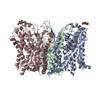
| Title | Complex of human ASCT2 with Syncytin-1 | |||||||||
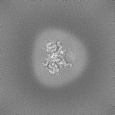
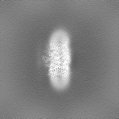
 Map data Map data | 2.62 Ang map of ASCT2-Syncytin-1. | |||||||||
 Sample Sample |
| |||||||||
 Keywords Keywords | Small neutral amino acid transporter / ASCT2 / Syncytin-1 /  Receptor binding domain / PROTEIN TRANSPORT Receptor binding domain / PROTEIN TRANSPORT | |||||||||
| Function / homology |  Function and homology information Function and homology informationsyncytium formation by plasma membrane fusion / glutamine secretion / syncytium formation / L-glutamine import across plasma membrane / glutamine transport / L-glutamine transmembrane transporter activity / L-serine transmembrane transporter activity / ligand-gated channel activity / neutral amino acid transport / amino acid transmembrane transporter activity ...syncytium formation by plasma membrane fusion / glutamine secretion / syncytium formation / L-glutamine import across plasma membrane / glutamine transport / L-glutamine transmembrane transporter activity / L-serine transmembrane transporter activity / ligand-gated channel activity / neutral amino acid transport / amino acid transmembrane transporter activity / Amino acid transport across the plasma membrane / neutral L-amino acid transmembrane transporter activity / L-aspartate transmembrane transporter activity / L-aspartate import across plasma membrane / symporter activity / myoblast fusion / amino acid transport / antiporter activity / RHOJ GTPase cycle / RHOQ GTPase cycle / protein homotrimerization / RHOH GTPase cycle / transport across blood-brain barrier / anatomical structure morphogenesis / RAC3 GTPase cycle / RAC1 GTPase cycle / basal plasma membrane / erythrocyte differentiation / melanosome / virus receptor activity / signaling receptor activity / extracellular exosome / membrane / metal ion binding / plasma membraneSimilarity search - Function | |||||||||
| Biological species |  Homo sapiens (human) Homo sapiens (human) | |||||||||
| Method | single particle reconstruction / cryo EM / Resolution: 2.62 Å | |||||||||
 Authors Authors | Khare S / Reyes N | |||||||||
| Funding support | European Union, 1 items
| |||||||||
 Citation Citation | Journal: Nat Struct Mol Biol / Year: 2024 Title: Receptor-recognition and antiviral mechanisms of retrovirus-derived human proteins. Authors: Shashank Khare / Miryam I Villalba / Juan C Canul-Tec / Arantza Balsebre Cajiao / Anand Kumar / Marija Backovic / Felix A Rey / Els Pardon / Jan Steyaert / Camilo Perez / Nicolas Reyes /     Abstract: Human syncytin-1 and suppressyn are cellular proteins of retroviral origin involved in cell-cell fusion events to establish the maternal-fetal interface in the placenta. In cell culture, they ...Human syncytin-1 and suppressyn are cellular proteins of retroviral origin involved in cell-cell fusion events to establish the maternal-fetal interface in the placenta. In cell culture, they restrict infections from members of the largest interference group of vertebrate retroviruses, and are regarded as host immunity factors expressed during development. At the core of the syncytin-1 and suppressyn functions are poorly understood mechanisms to recognize a common cellular receptor, the membrane transporter ASCT2. Here, we present cryo-electron microscopy structures of human ASCT2 in complexes with the receptor-binding domains of syncytin-1 and suppressyn. Despite their evolutionary divergence, the two placental proteins occupy similar positions in ASCT2, and are stabilized by the formation of a hybrid β-sheet or 'clamp' with the receptor. Structural predictions of the receptor-binding domains of extant retroviruses indicate overlapping binding interfaces and clamping sites with ASCT2, revealing a competition mechanism between the placental proteins and the retroviruses. Our work uncovers a common ASCT2 recognition mechanism by a large group of endogenous and disease-causing retroviruses, and provides high-resolution views on how placental human proteins exert morphological and immunological functions. | |||||||||
| History |
|
- Structure visualization
| Supplemental images |
|---|

- Downloads & links
-EMDB archive
| Map data | emd_17192.map.gz | 122.1 MB | EMDB map data format | |
|---|---|---|---|---|
| Header (meta data) | emd-17192-v30.xmlemd-17192.xml | 22.1 KB 22.1 KB | Display Display | EMDB header |
| FSC (resolution estimation) | emd_17192_fsc.xml | 13.2 KB | Display | FSC data file |
| Images |  emd_17192.png emd_17192.png | 116.8 KB | ||
| Filedesc metadata | emd-17192.cif.gz | 7 KB | ||
| Others | emd_17192_half_map_1.map.gzemd_17192_half_map_2.map.gz | 226.8 MB 226.8 MB | ||
| Archive directory |  http://ftp.pdbj.org/pub/emdb/structures/EMD-17192ftp://ftp.pdbj.org/pub/emdb/structures/EMD-17192 http://ftp.pdbj.org/pub/emdb/structures/EMD-17192ftp://ftp.pdbj.org/pub/emdb/structures/EMD-17192 | HTTPS FTP |
-Related structure data
| Related structure data |  8ouhMC  8oudC 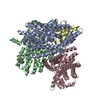 8ouiC  8oujC M: atomic model generated by this map C: citing same article ( |
|---|---|
| Similar structure data |

-Links
| EMDB pages | EMDB (EBI/PDBe) / EMDataResource |
|---|---|
| Related items in Molecule of the Month |


-Map
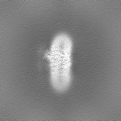
| File | Download / File: emd_17192.map.gz / Format: CCP4 / Size: 244.1 MB / Type: IMAGE STORED AS FLOATING POINT NUMBER (4 BYTES) | ||||||||||||||||||||||||||||||||||||
|---|---|---|---|---|---|---|---|---|---|---|---|---|---|---|---|---|---|---|---|---|---|---|---|---|---|---|---|---|---|---|---|---|---|---|---|---|---|
| Annotation | 2.62 Ang map of ASCT2-Syncytin-1. | ||||||||||||||||||||||||||||||||||||
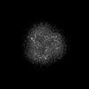
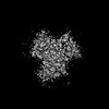
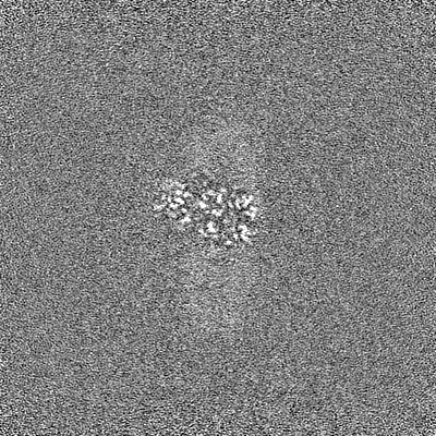
| Projections & slices | Image control
Images are generated by Spider. | ||||||||||||||||||||||||||||||||||||
| Voxel size | X=Y=Z: 0.731 Å | ||||||||||||||||||||||||||||||||||||
| Density |
| ||||||||||||||||||||||||||||||||||||
| Symmetry | Space group: 1 | ||||||||||||||||||||||||||||||||||||
| Details | EMDB XML:
|
 Z (Sec.)
 Y (Row.)
 X (Col.)


-Supplemental data
-Half map: #2
| File | emd_17192_half_map_1.map | ||||||||||||
|---|---|---|---|---|---|---|---|---|---|---|---|---|---|
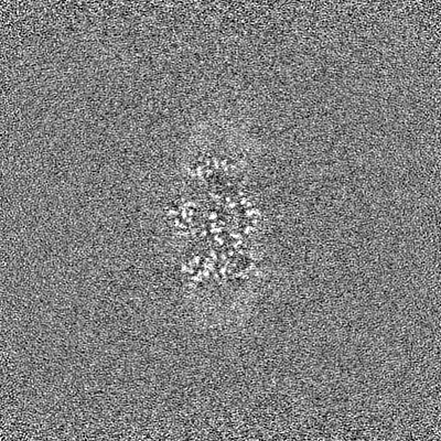
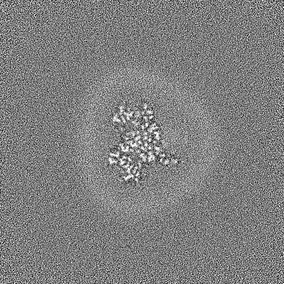
| Projections & Slices |
| ||||||||||||
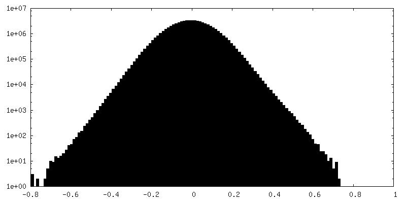
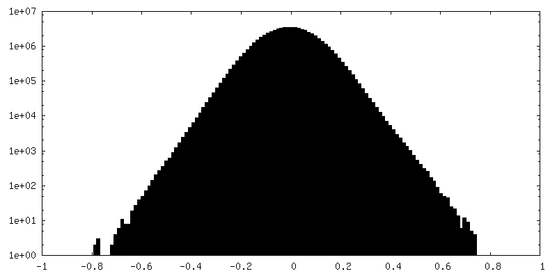
| Density Histograms |


-Half map: #1
| File | emd_17192_half_map_2.map | ||||||||||||
|---|---|---|---|---|---|---|---|---|---|---|---|---|---|
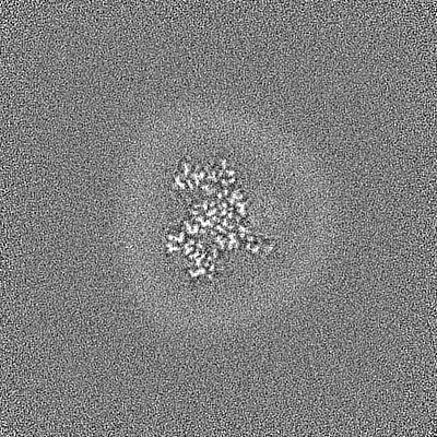
| Projections & Slices |
| ||||||||||||
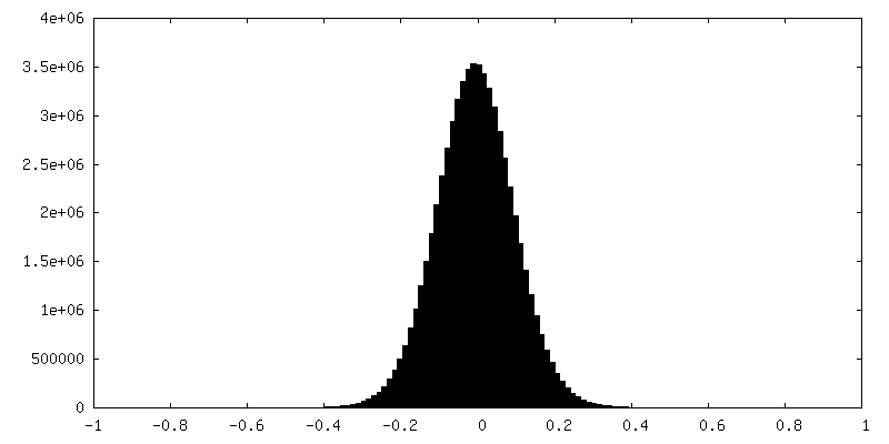
| Density Histograms |


- Sample components
-Entire : Complex of ASCT2 with Synctin-1
| Entire | Name: Complex of ASCT2 with Synctin-1 |
|---|---|
| Components |
|
-Supramolecule #1: Complex of ASCT2 with Synctin-1
| Supramolecule | Name: Complex of ASCT2 with Synctin-1 / type: complex / ID: 1 / Parent: 0 / Macromolecule list: #1-#2 |
|---|---|
| Molecular weight | Theoretical: 180 kDa/nm |
-Supramolecule #2: Alanine Serine Cysteine Transporter 2
| Supramolecule | Name: Alanine Serine Cysteine Transporter 2 / type: complex / ID: 2 / Parent: 1 / Macromolecule list: #1 |
|---|---|
| Source (natural) | Organism: Homo sapiens (human) |
-Supramolecule #3: Syncytin-1
| Supramolecule | Name: Syncytin-1 / type: complex / ID: 3 / Parent: 1 / Macromolecule list: #2 |
|---|---|
| Source (natural) | Organism: Homo sapiens (human) |
-Macromolecule #1: Neutral amino acid transporter B(0)
| Macromolecule | Name: Neutral amino acid transporter B(0) / type: protein_or_peptide / ID: 1 / Number of copies: 3 / Enantiomer: LEVO |
|---|---|
| Source (natural) | Organism: Homo sapiens (human) |
| Molecular weight | Theoretical: 58.984566 KDa |
| Recombinant expression | Organism: Homo sapiens (human) |
| Sequence | String: MWSHPQFEKS SGGLEVLFQG PMVADPPRDS KGLAAAEPTA NGGLALASIE DQGAAAGGYC GSRDQVRRCL RANLLVLLTV VAVVAGVAL GLGVSGAGGA LALGPERLSA FVFPGELLLR LLRMIILPLV VCSLIGGAAS LDPGALGRLG AWALLFFLVT T LLASALGV ...String: MWSHPQFEKS SGGLEVLFQG PMVADPPRDS KGLAAAEPTA NGGLALASIE DQGAAAGGYC GSRDQVRRCL RANLLVLLTV VAVVAGVAL GLGVSGAGGA LALGPERLSA FVFPGELLLR LLRMIILPLV VCSLIGGAAS LDPGALGRLG AWALLFFLVT T LLASALGV GLALALQPGA ASAAINASVG AAGSAENAPS KEVLDSFLDL ARNIFPSNLV SAAFRSYSTT YEERNITGTR VK VPVGQEV EGMNILGLVV FAIVFGVALR KLGPEGELLI RFFNSFNEAT MVLVSWIMWY APVGIMFLVA GKIVEMEDVG LLF ARLGKY ILCCLLGHAI HGLLVLPLIY FLFTRKNPYR FLWGIVTPLA TAFGTSSSSA TLPLMMKCVE ENNGVAKHIS RFIL PIGAT VNMDGAALFQ CVAAVFIAQL SQQSLDFVKI ITILVTATAS SVGAAGIPAG GVLTLAIILE AVNLPVDHIS LILAV DWLV DRSCTVLNVE GDALGAGLLQ NYVDRTESRS TEPELIQVKS ELPLDPLPVP TEEGNPLLKH YRGPAGDATV ASEKES VM UniProtKB: Neutral amino acid transporter B(0) |
-Macromolecule #2: Syncytin-1
| Macromolecule | Name: Syncytin-1 / type: protein_or_peptide / ID: 2 / Number of copies: 1 / Enantiomer: LEVO |
|---|---|
| Source (natural) | Organism: Homo sapiens (human) |
| Molecular weight | Theoretical: 49.24593 KDa |
| Recombinant expression | Organism:  Drosophila melanogaster (fruit fly) Drosophila melanogaster (fruit fly) |
| Sequence | String: AVVAFVGLSL GAPPPCRCMT SSSPYQEFLW RMQRPGNIDA PSYRSLSKGT PTFTAHTHMP RNCYHSATLC MHANTHYWTG KMINPSCPG GLGVTVCWTY FTQTGMSDGG GVQDQAREKH VKEVISQLTR VHGTSSPYKG LDLSKLHETL RTHTRLVSLF N TTLTGLHE ...String: AVVAFVGLSL GAPPPCRCMT SSSPYQEFLW RMQRPGNIDA PSYRSLSKGT PTFTAHTHMP RNCYHSATLC MHANTHYWTG KMINPSCPG GLGVTVCWTY FTQTGMSDGG GVQDQAREKH VKEVISQLTR VHGTSSPYKG LDLSKLHETL RTHTRLVSLF N TTLTGLHE VSAQNPTNSW ICLPLNFRPY VSIPVPEQWN NFSTEINTTS VLVGPLVSNL EITHTSNLTC VKFSNTTYTT NS QCIRWVT PPTQIVCLPS GIFFVCGTSA YRCLNGSSES MCFLSFLVPP MTIYTEQDLY NYVISKPRNK RVPILPFVIG AGV LGALGT GIGGITTSTQ FYYKLSQELN GDMERVADSL VTLQDQLNSL AAVVLQNRRA LDLLTAERGG TCLFLGEECC YYVN QSGIV TEKVKEIRDR IQRRAEELRN TGPWGSGLEV LFQGPGPEPE A UniProtKB: Syncytin-1 |
-Macromolecule #3: ALANINE
| Macromolecule | Name: ALANINE / type: ligand / ID: 3 / Number of copies: 2 / Formula: ALA |
|---|---|
| Molecular weight | Theoretical: 89.093 Da |
| Chemical component information |  ChemComp-ALA: |
-Experimental details
-Structure determination
| Method | cryo EM |
|---|---|
 Processing Processing | single particle reconstruction |
| Aggregation state | particle |
-Sample preparation
| Concentration | 9 mg/mL | ||||||||||||||||||
|---|---|---|---|---|---|---|---|---|---|---|---|---|---|---|---|---|---|---|---|
| Buffer | pH: 7.4 Component:
| ||||||||||||||||||
| Grid | Model: Quantifoil R1.2/1.3 / Material: GOLD / Mesh: 300 / Support film - Material: CARBON / Support film - topology: CONTINUOUS / Support film - Film thickness: 10 / Pretreatment - Type: GLOW DISCHARGE / Pretreatment - Time: 40 sec. / Pretreatment - Atmosphere: AIR / Pretreatment - Pressure: 0.003 kPa | ||||||||||||||||||
| Vitrification | Cryogen name: ETHANE / Chamber humidity: 100 % / Chamber temperature: 277 K / Instrument: FEI VITROBOT MARK IV |
- Electron microscopy
| Microscope | FEI TITAN KRIOS |
|---|---|
| Electron beam | Acceleration voltage: 300 kV / Electron source: FIELD EMISSION GUN |
| Electron optics | C2 aperture diameter: 50.0 µm / Illumination mode: FLOOD BEAM / Imaging mode: BRIGHT FIELDBright-field microscopy / Cs: 2.7 mm / Nominal defocus max: 1.5 µm / Nominal defocus min: 0.4 µm / Nominal magnification: 165000 |
| Specialist optics | Phase plate: VOLTA PHASE PLATE / Energy filter - Name: TFS Selectris X / Energy filter - Slit width: 10 eV |
| Sample stage | Cooling holder cryogen: NITROGEN |
| Software | Name: EPU (ver. 2) |
| Image recording | Film or detector model: FEI FALCON IV (4k x 4k) / Number grids imaged: 1 / Number real images: 15376 / Average exposure time: 6.0 sec. / Average electron dose: 53.4 e/Å2 |
| Experimental equipment |  Model: Titan Krios / Image courtesy: FEI Company |
-Image processing
| Particle selection | Number selected: 4018074 |
|---|---|
| Startup model | Type of model: PDB ENTRY PDB model - PDB ID: |
| Initial angle assignment | Type: MAXIMUM LIKELIHOOD / Software - Name: cryoSPARC (ver. 3.3) |
| Final angle assignment | Type: MAXIMUM LIKELIHOOD / Details: Need to ask |
| Final reconstruction | Algorithm: FOURIER SPACE / Resolution.type: BY AUTHOR / Resolution: 2.62 Å / Resolution method: FSC 0.143 CUT-OFF / Software - Name: cryoSPARC (ver. 3.3) / Number images used: 136195 |
| FSC plot (resolution estimation) |  |
-Atomic model buiding 1
| Initial model |
| |||||||||
|---|---|---|---|---|---|---|---|---|---|---|
| Software | Name: Coot (ver. 0.9.8.3) | |||||||||
| Refinement | Space: REAL / Protocol: FLEXIBLE FIT / Overall B value: 60.1 | |||||||||
| Output model | PDB-8ouh: |

