 Movie
Movie Controller


+ Open data

- Basic information
| Entry | Database: PDB / ID: 8ohi | ||||||
|---|---|---|---|---|---|---|---|
| Title | Structure of the Fmoc-Tau-PAM4 Type 2 amyloid fibril | ||||||
 Components Components | Microtubule-associated protein tau Tau protein Tau protein | ||||||
 Keywords Keywords | PROTEIN FIBRIL / amyloid / tau / helical / cross-beta / fibril / neurodegeneration / Fmoc | ||||||
| Function / homology |  Function and homology information Function and homology informationplus-end-directed organelle transport along microtubule / axonal transport / histone-dependent DNA binding / neurofibrillary tangle assembly / positive regulation of diacylglycerol kinase activity / negative regulation of establishment of protein localization to mitochondrion / neurofibrillary tangle / positive regulation of protein localization to synapse / microtubule lateral binding / tubulin complex ...plus-end-directed organelle transport along microtubule / axonal transport / histone-dependent DNA binding / neurofibrillary tangle assembly / positive regulation of diacylglycerol kinase activity / negative regulation of establishment of protein localization to mitochondrion / neurofibrillary tangle / positive regulation of protein localization to synapse / microtubule lateral binding / tubulin complex / phosphatidylinositol bisphosphate binding / main axon / regulation of long-term synaptic depression / negative regulation of kinase activity / negative regulation of tubulin deacetylation / generation of neurons / regulation of chromosome organization / positive regulation of protein localization / rRNA metabolic process / internal protein amino acid acetylation / regulation of mitochondrial fission / lipoprotein particle binding / intracellular distribution of mitochondria / axonal transport of mitochondrion / axon development / central nervous system neuron development / regulation of microtubule polymerization / microtubule polymerization / minor groove of adenine-thymine-rich DNA binding / negative regulation of mitochondrial membrane potential / dynactin binding / glial cell projection / apolipoprotein binding / protein polymerization / negative regulation of mitochondrial fission / axolemma / Caspase-mediated cleavage of cytoskeletal proteins / regulation of microtubule polymerization or depolymerization / positive regulation of axon extension / supramolecular fiber organization / Activation of AMPK downstream of NMDARs / regulation of microtubule cytoskeleton organization / stress granule assembly / cytoplasmic microtubule organization / regulation of cellular response to heat / regulation of calcium-mediated signaling / axon cytoplasm / positive regulation of microtubule polymerization / cellular response to brain-derived neurotrophic factor stimulus / somatodendritic compartment / synapse assembly / phosphatidylinositol binding / nuclear periphery / cellular response to nerve growth factor stimulus / positive regulation of superoxide anion generation / protein phosphatase 2A binding / regulation of autophagy / astrocyte activation / synapse organization / microglial cell activation / response to lead ion / Hsp90 protein binding / regulation of synaptic plasticity / PKR-mediated signaling / protein homooligomerization / cytoplasmic ribonucleoprotein granule / memory / microtubule cytoskeleton organization / cellular response to reactive oxygen species / SH3 domain binding / neuron projection development / activation of cysteine-type endopeptidase activity involved in apoptotic process / microtubule cytoskeleton / protein-macromolecule adaptor activity / single-stranded DNA binding / cell-cell signaling / cellular response to heat / cell body / actin binding / growth cone / protein-folding chaperone binding / double-stranded DNA binding / microtubule binding / microtubule / amyloid fibril formation / sequence-specific DNA binding / dendritic spine / learning or memory / neuron projection / nuclear speck / membrane raft / axon / negative regulation of gene expression / dendrite / neuronal cell body / DNA damage response / protein kinase binding / enzyme binding / mitochondrion / DNA bindingSimilarity search - Function | ||||||
| Biological species | synthetic construct (others) | ||||||
| Method | ELECTRON MICROSCOPY / helical reconstruction / cryo EM / Resolution: 2.8 Å | ||||||
 Authors Authors | Wilkinson, M. / Louros, N. / Tsaka, G. / Ramakers, M. / Morelli, C. / Garcia, T. / Gallardo, R.U. / D'Haeyer, S. / Goossens, V. / Audenaert, D. ...Wilkinson, M. / Louros, N. / Tsaka, G. / Ramakers, M. / Morelli, C. / Garcia, T. / Gallardo, R.U. / D'Haeyer, S. / Goossens, V. / Audenaert, D. / Thal, D.R. / Ranson, N.A. / Radford, S.E. / Rousseau, F. / Schymkowitz, J. | ||||||
| Funding support |  Belgium, 1items Belgium, 1items
| ||||||
 Citation Citation | Journal: Nat Commun / Year: 2024 Title: Local structural preferences in shaping tau amyloid polymorphism. Authors: Nikolaos Louros / Martin Wilkinson / Grigoria Tsaka / Meine Ramakers / Chiara Morelli / Teresa Garcia / Rodrigo Gallardo / Sam D'Haeyer / Vera Goossens / Dominique Audenaert / Dietmar Rudolf ...Authors: Nikolaos Louros / Martin Wilkinson / Grigoria Tsaka / Meine Ramakers / Chiara Morelli / Teresa Garcia / Rodrigo Gallardo / Sam D'Haeyer / Vera Goossens / Dominique Audenaert / Dietmar Rudolf Thal / Ian R Mackenzie / Rosa Rademakers / Neil A Ranson / Sheena E Radford / Frederic Rousseau / Joost Schymkowitz /   Abstract: Tauopathies encompass a group of neurodegenerative disorders characterised by diverse tau amyloid fibril structures. The persistence of polymorphism across tauopathies suggests that distinct ...Tauopathies encompass a group of neurodegenerative disorders characterised by diverse tau amyloid fibril structures. The persistence of polymorphism across tauopathies suggests that distinct pathological conditions dictate the adopted polymorph for each disease. However, the extent to which intrinsic structural tendencies of tau amyloid cores contribute to fibril polymorphism remains uncertain. Using a combination of experimental approaches, we here identify a new amyloidogenic motif, PAM4 (Polymorphic Amyloid Motif of Repeat 4), as a significant contributor to tau polymorphism. Calculation of per-residue contributions to the stability of the fibril cores of different pathologic tau structures suggests that PAM4 plays a central role in preserving structural integrity across amyloid polymorphs. Consistent with this, cryo-EM structural analysis of fibrils formed from a synthetic PAM4 peptide shows that the sequence adopts alternative structures that closely correspond to distinct disease-associated tau strains. Furthermore, in-cell experiments revealed that PAM4 deletion hampers the cellular seeding efficiency of tau aggregates extracted from Alzheimer's disease, corticobasal degeneration, and progressive supranuclear palsy patients, underscoring PAM4's pivotal role in these tauopathies. Together, our results highlight the importance of the intrinsic structural propensity of amyloid core segments to determine the structure of tau in cells, and in propagating amyloid structures in disease. | ||||||
| History |
|
- Structure visualization
| Structure viewer | Molecule: MolmilJmol/JSmol |
|---|
- Downloads & links
-Download
| PDBx/mmCIF format | 8ohi.cif.gz | 55.8 KB | Display | PDBx/mmCIF format |
|---|---|---|---|---|
| PDB format | pdb8ohi.ent.gz | 44.9 KB | Display | PDB format |
| PDBx/mmJSON format | 8ohi.json.gz | Tree view | PDBx/mmJSON format | |
| Others |  Other downloads Other downloads |
-Validation report
| Arichive directory | https://data.pdbj.org/pub/pdb/validation_reports/oh/8ohiftp://data.pdbj.org/pub/pdb/validation_reports/oh/8ohi | HTTPS FTP |
|---|
-Related structure data
| Related structure data |  16881MC 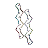 8oh2C 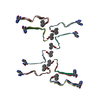 8ohpC 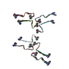 8oi0C C: citing same article ( M: map data used to model this data |
|---|---|
| Similar structure data |


-Links
 PDBj
PDBj


- Assembly
| Deposited unit | 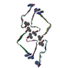
|
|---|---|
| 1 |
|
-Components
| #1: Protein/peptide | Tau protein / Neurofibrillary tangle protein / Paired helical filament-tau / PHF-tau Mass: 1652.269 Da / Num. of mol.: 18 / Source method: obtained synthetically Details: 13-residue peptide of the PAM4 motif of Tau, corresponding to residues 350-362 of the Tau repeat domain. The peptide is supposed to be N-terminally acetylated but 10% (by mass spec) was ...Details: 13-residue peptide of the PAM4 motif of Tau, corresponding to residues 350-362 of the Tau repeat domain. The peptide is supposed to be N-terminally acetylated but 10% (by mass spec) was still adducted to the Fmoc protection group from synthesis. The peptide in the fibrils is dominated by this impurity. The peptide is also C-terminally amidated. Source: (synth.) synthetic construct (others) / References: UniProt: P10636 Has ligand of interest | Y | |
|---|
-Experimental details
-Experiment
| Experiment | Method: ELECTRON MICROSCOPY |
|---|---|
| EM experiment | Aggregation state: FILAMENT / 3D reconstruction method: helical reconstruction |
- Sample preparation
| Component | Name: Amyloid fibril form 1a of Tau-PAM4 peptide adducted with the Fmoc protection group Type: COMPLEX / Details: Synthesised peptide assembled into amyloid fibril / Entity ID: all / Source: NATURAL |
|---|---|
| Molecular weight | Experimental value: NO |
| Source (natural) | Organism: synthetic construct (others) |
| Buffer solution | pH: 7 Details: Peptide resuspended in MilliQ water for aggregation reaction |
| Specimen | Conc.: 0.6 mg/ml / Embedding applied: NO / Shadowing applied: NO / Staining applied: NO / Vitrification applied: YES |
| Specimen support | Grid material: COPPER / Grid mesh size: 300 divisions/in. / Grid type: EMS Lacey Carbon |
| Vitrification | Instrument: FEI VITROBOT MARK IV / Cryogen name: ETHANE / Humidity: 90 % / Chamber temperature: 279 K |
- Electron microscopy imaging
| Experimental equipment |  Model: Titan Krios / Image courtesy: FEI Company |
|---|---|
| Microscopy | Model: FEI TITAN KRIOS |
| Electron gun | Electron source: FIELD EMISSION GUN / Accelerating voltage: 300 kV / Illumination mode: FLOOD BEAM |
| Electron lens | Mode: BRIGHT FIELDBright-field microscopy / Nominal magnification: 130000 X / Nominal defocus max: 2400 nm / Nominal defocus min: 1200 nm / Cs: 2.7 mm / C2 aperture diameter: 50 µm / Alignment procedure: COMA FREE |
| Specimen holder | Specimen holder model: FEI TITAN KRIOS AUTOGRID HOLDER |
| Image recording | Average exposure time: 5 sec. / Electron dose: 32 e/Å2 / Film or detector model: FEI FALCON IV (4k x 4k) / Num. of grids imaged: 1 / Num. of real images: 1957 Details: Movies were collected as 1204 EER frames compressed and re-grouped into 35 TIF fractions |
| EM imaging optics | Energyfilter name: TFS Selectris / Energyfilter slit width: 10 eV |
| Image scans | Width: 4096 / Height: 4096 |
- Processing
| EM software |
| ||||||||||||||||||||||||||||||||||||||||
|---|---|---|---|---|---|---|---|---|---|---|---|---|---|---|---|---|---|---|---|---|---|---|---|---|---|---|---|---|---|---|---|---|---|---|---|---|---|---|---|---|---|
| CTF correction | Type: PHASE FLIPPING AND AMPLITUDE CORRECTION | ||||||||||||||||||||||||||||||||||||||||
| Helical symmerty | Angular rotation/subunit: -1.45 ° / Axial rise/subunit: 4.8 Å / Axial symmetry: C1 | ||||||||||||||||||||||||||||||||||||||||
| Particle selection | Num. of particles selected: 520390 | ||||||||||||||||||||||||||||||||||||||||
| 3D reconstruction | Resolution: 2.8 Å / Resolution method: FSC 0.143 CUT-OFF / Num. of particles: 11255 / Symmetry type: HELICAL | ||||||||||||||||||||||||||||||||||||||||
| Atomic model building | B value: 48 / Protocol: AB INITIO MODEL / Space: REAL / Target criteria: Cross-correlation coefficient Details: Initial model built manually in coot, no starting template | ||||||||||||||||||||||||||||||||||||||||
| Refine LS restraints |
|
