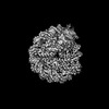
 Movie
Movie Controller


+ Open data

- Basic information
| Entry | Database: PDB / ID: 8of4 | ||||||
|---|---|---|---|---|---|---|---|
| Title | Nucleosome Bound human SIRT6 (Composite) | ||||||
 Components Components |
| ||||||
 Keywords Keywords |  GENE REGULATION / Transferase / Deacetylase / Histone H3 deacetylation GENE REGULATION / Transferase / Deacetylase / Histone H3 deacetylation | ||||||
| Function / homology |  Function and homology information Function and homology informationNAD-dependent histone H3K56 deacetylase activity / NAD-dependent histone H3K18 deacetylase activity / ketone biosynthetic process / NAD-dependent histone H3K9 deacetylase activity / protein delipidation / regulation of lipid catabolic process / NAD+- protein-lysine ADP-ribosyltransferase activity / NAD-dependent protein demyristoylase activity / NAD-dependent protein depalmitoylase activity / chromosome, subtelomeric region ...NAD-dependent histone H3K56 deacetylase activity / NAD-dependent histone H3K18 deacetylase activity / ketone biosynthetic process / NAD-dependent histone H3K9 deacetylase activity / protein delipidation / regulation of lipid catabolic process / NAD+- protein-lysine ADP-ribosyltransferase activity / NAD-dependent protein demyristoylase activity / NAD-dependent protein depalmitoylase activity / chromosome, subtelomeric region / pericentric heterochromatin formation / NAD+-protein-arginine ADP-ribosyltransferase activity / positive regulation of protein localization to chromatin / DNA damage sensor activity / positive regulation of stem cell differentiation / positive regulation of blood vessel branching / NAD-dependent protein lysine deacetylase activity / protein localization to site of double-strand break / retrotransposon silencing / protein acetyllysine N-acetyltransferase / cardiac muscle cell differentiation / NAD-dependent histone deacetylase activity / positive regulation of chondrocyte proliferation / positive regulation of telomere maintenance / protein deacetylation / negative regulation of glucose import / TORC2 complex binding / lncRNA binding / negative regulation of glycolytic process / negative regulation of protein localization to chromatin / DNA repair-dependent chromatin remodeling / positive regulation of double-strand break repair / positive regulation of vascular endothelial cell proliferation / negative regulation of gene expression, epigenetic / regulation of double-strand break repair via homologous recombination / negative regulation of protein import into nucleus / positive regulation of stem cell population maintenance / positive regulation of stem cell proliferation / regulation of protein secretion / negative regulation of transcription elongation by RNA polymerase II / site of DNA damage / NAD+-protein ADP-ribosyltransferase activity / negative regulation of cellular senescence / subtelomeric heterochromatin formation / regulation of lipid metabolic process / NAD+ ADP-ribosyltransferase activity / Transferases; Glycosyltransferases; Pentosyltransferases / nucleosome binding / NAD+ binding / positive regulation of fat cell differentiation / negative regulation of gluconeogenesis / pericentric heterochromatin / regulation of protein localization to plasma membrane / response to UV / Transferases; Acyltransferases; Transferring groups other than aminoacyl groups / nucleotidyltransferase activity / positive regulation of protein export from nucleus / determination of adult lifespan / circadian regulation of gene expression / base-excision repair / protein destabilization / regulation of circadian rhythm / positive regulation of insulin secretion / chromatin DNA binding / Pre-NOTCH Transcription and Translation / structural constituent of chromatin / transcription corepressor activity / positive regulation of fibroblast proliferation / double-strand break repair / nucleosome / positive regulation of proteasomal ubiquitin-dependent protein catabolic process / glucose homeostasis / site of double-strand break / positive regulation of cold-induced thermogenesis / Processing of DNA double-strand break ends / damaged DNA binding / chromatin remodeling / protein heterodimerization activity / negative regulation of cell population proliferation / intracellular membrane-bounded organelle / chromatin binding / chromatin / negative regulation of transcription by RNA polymerase II / endoplasmic reticulum / protein homodimerization activity / DNA binding / zinc ion binding / nucleoplasm / nucleusSimilarity search - Function | ||||||
| Biological species | Xenopus laevis (African clawed frog) Homo sapiens (human) Homo sapiens (human) | ||||||
| Method | ELECTRON MICROSCOPY / single particle reconstruction / cryo EM / Resolution: 2.94 Å | ||||||
 Authors Authors | Smirnova, E. / Bignon, E. / Schultz, P. / Papai, G. / Ben-Shem, A. | ||||||
| Funding support |  France, 1items France, 1items
| ||||||
 Citation Citation | Journal: Elife / Year: 2024 Title: Binding to nucleosome poises human SIRT6 for histone H3 deacetylation. Authors: Ekaterina Smirnova / Emmanuelle Bignon / Patrick Schultz / Gabor Papai / Adam Ben Shem / Abstract: Sirtuin 6 (SIRT6) is an NAD-dependent histone H3 deacetylase that is prominently found associated with chromatin, attenuates transcriptionally active promoters and regulates DNA repair, metabolic ...Sirtuin 6 (SIRT6) is an NAD-dependent histone H3 deacetylase that is prominently found associated with chromatin, attenuates transcriptionally active promoters and regulates DNA repair, metabolic homeostasis and lifespan. Unlike other sirtuins, it has low affinity to free histone tails but demonstrates strong binding to nucleosomes. It is poorly understood how SIRT6 docking on nucleosomes stimulates its histone deacetylation activity. Here, we present the structure of human SIRT6 bound to a nucleosome determined by cryogenic electron microscopy. The zinc finger domain of SIRT6 associates tightly with the acidic patch of the nucleosome through multiple arginine anchors. The Rossmann fold domain binds to the terminus of the looser DNA half of the nucleosome, detaching two turns of the DNA from the histone octamer and placing the NAD binding pocket close to the DNA exit site. This domain shows flexibility with respect to the fixed zinc finger and moves with, but also relative to, the unwrapped DNA terminus. We apply molecular dynamics simulations of the histone tails in the nucleosome to show that in this mode of interaction, the active site of SIRT6 is perfectly poised to catalyze deacetylation of the H3 histone tail and that the partial unwrapping of the DNA allows even lysines close to the H3 core to reach the enzyme. #1: Journal: Elife / Year: 2023Title: Binding to nucleosome poises SIRT6 for histone H3 de-acetylation Authors: Smirnova, E. / Bignon, E. / Schultz, P. / Papai, G. / Ben-Shem, A. | ||||||
| History |
|
- Structure visualization
| Structure viewer | Molecule: MolmilJmol/JSmol |
|---|
- Downloads & links
-Download
| PDBx/mmCIF format | 8of4.cif.gz | 358 KB | Display | PDBx/mmCIF format |
|---|---|---|---|---|
| PDB format | pdb8of4.ent.gz | 265.8 KB | Display | PDB format |
| PDBx/mmJSON format | 8of4.json.gz | Tree view | PDBx/mmJSON format | |
| Others |  Other downloads Other downloads |
-Validation report
| Arichive directory | https://data.pdbj.org/pub/pdb/validation_reports/of/8of4ftp://data.pdbj.org/pub/pdb/validation_reports/of/8of4 | HTTPS FTP |
|---|
-Related structure data
| Related structure data |  16845MC C: citing same article ( M: map data used to model this data |
|---|---|
| Similar structure data |


-Links
 PDBj
PDBj


- Assembly
| Deposited unit | 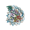
|
|---|---|
| 1 |
|
-Components
-Protein , 5 types, 9 molecules AEBFCGDHL
| #1: Protein | Mass: 15421.101 Da / Num. of mol.: 2 Source method: isolated from a genetically manipulated source Source: (gene. exp.) Xenopus laevis (African clawed frog) / Production host:  Escherichia coli (E. coli) / References: UniProt: P84233 Escherichia coli (E. coli) / References: UniProt: P84233#2: Protein | Mass: 11394.426 Da / Num. of mol.: 2 Source method: isolated from a genetically manipulated source Source: (gene. exp.) Xenopus laevis (African clawed frog) / Production host: Escherichia coli (E. coli) / References: UniProt: P62799#3: Protein | Mass: 14093.436 Da / Num. of mol.: 2 Source method: isolated from a genetically manipulated source Source: (gene. exp.) Xenopus laevis (African clawed frog) / Production host: Escherichia coli (E. coli) / References: UniProt: P06897#4: Protein | Mass: 13979.291 Da / Num. of mol.: 2 Source method: isolated from a genetically manipulated source Source: (gene. exp.) Xenopus laevis (African clawed frog) / Gene: LOC108704303 / Production host: Escherichia coli (E. coli) / References: UniProt: A0A8J0U496#7: Protein | | Mass: 39180.914 Da / Num. of mol.: 1 Source method: isolated from a genetically manipulated source Source: (gene. exp.) Homo sapiens (human) / Gene: SIRT6, SIR2L6 / Production host: Escherichia coli (E. coli)References: UniProt: Q8N6T7, Transferases; Acyltransferases; Transferring groups other than aminoacyl groups, protein acetyllysine N-acetyltransferase, Transferases; Glycosyltransferases; Pentosyltransferases |
|---|
-DNA chain , 2 types, 2 molecules IJ
| #5: DNA chain | Mass: 44520.383 Da / Num. of mol.: 1 Source method: isolated from a genetically manipulated source Source: (gene. exp.) Xenopus laevis (African clawed frog) / Production host: Escherichia coli (E. coli) |
|---|---|
| #6: DNA chain | Mass: 44991.660 Da / Num. of mol.: 1 Source method: isolated from a genetically manipulated source Source: (gene. exp.) Xenopus laevis (African clawed frog) / Production host: Escherichia coli (E. coli) |
-Non-polymers , 1 types, 1 molecules 
| #8: Chemical | ChemComp-ZN /  Mass: 65.409 Da / Num. of mol.: 1 / Source method: obtained synthetically / Formula: Zn / Feature type: SUBJECT OF INVESTIGATION Mass: 65.409 Da / Num. of mol.: 1 / Source method: obtained synthetically / Formula: Zn / Feature type: SUBJECT OF INVESTIGATION |
|---|
-Details
| Has ligand of interest | Y |
|---|
-Experimental details
-Experiment
| Experiment | Method: ELECTRON MICROSCOPY |
|---|---|
| EM experiment | Aggregation state: PARTICLE / 3D reconstruction method: single particle reconstruction |
- Sample preparation
| Component |
| ||||||||||||||||||
|---|---|---|---|---|---|---|---|---|---|---|---|---|---|---|---|---|---|---|---|
| Molecular weight |
| ||||||||||||||||||
| Source (natural) |
| ||||||||||||||||||
| Source (recombinant) |
| ||||||||||||||||||
| Buffer solution | pH: 7.5 | ||||||||||||||||||
| Specimen | Embedding applied: NO / Shadowing applied: NO / Staining applied: NO / Vitrification applied: YES | ||||||||||||||||||
| Specimen support | Grid material: COPPER/RHODIUM / Grid mesh size: 300 divisions/in. / Grid type: Quantifoil R3.5/1 | ||||||||||||||||||
| Vitrification | Cryogen name: ETHANE |
- Electron microscopy imaging
| Experimental equipment |  Model: Titan Krios / Image courtesy: FEI Company | ||||||||||||
|---|---|---|---|---|---|---|---|---|---|---|---|---|---|
| EM imaging | Accelerating voltage: 300 kV / C2 aperture diameter: 50 µm / Cryogen: NITROGEN / Electron source
| ||||||||||||
| Image recording |
| ||||||||||||
| EM imaging optics |
|
- Processing
| EM software |
| ||||||||||||||||||||||||||||||||||||||||||||||||
|---|---|---|---|---|---|---|---|---|---|---|---|---|---|---|---|---|---|---|---|---|---|---|---|---|---|---|---|---|---|---|---|---|---|---|---|---|---|---|---|---|---|---|---|---|---|---|---|---|---|
| CTF correction | Type: PHASE FLIPPING AND AMPLITUDE CORRECTION | ||||||||||||||||||||||||||||||||||||||||||||||||
| Particle selection | Num. of particles selected: 2033169 | ||||||||||||||||||||||||||||||||||||||||||||||||
| 3D reconstruction | Resolution: 2.94 Å / Resolution method: FSC 0.143 CUT-OFF / Num. of particles: 439796 / Algorithm: BACK PROJECTION / Symmetry type: POINT | ||||||||||||||||||||||||||||||||||||||||||||||||
| Atomic model building |
|


