 Movie
Movie Controller

[English] 日本語
 Yorodumi
- PDB-8kcm: MmCPDII-DNA complex containing low-dosage, light induced repaired DNA. -
+ Open data

- Basic information
| Entry | Database: PDB / ID: 8kcm | ||||||||||||
|---|---|---|---|---|---|---|---|---|---|---|---|---|---|
| Title | MmCPDII-DNA complex containing low-dosage, light induced repaired DNA. | ||||||||||||
 Components Components |
| ||||||||||||
 Keywords Keywords |  DNA BINDING PROTEIN / Flavoprotein / photolyase / light driven electron transfer / DNA repair / time-resolved serial crystallography. DNA BINDING PROTEIN / Flavoprotein / photolyase / light driven electron transfer / DNA repair / time-resolved serial crystallography. | ||||||||||||
| Function / homology |  Function and homology informationdeoxyribodipyrimidine photo-lyase / deoxyribodipyrimidine photo-lyase activity / DNA repair / DNA binding Function and homology informationdeoxyribodipyrimidine photo-lyase / deoxyribodipyrimidine photo-lyase activity / DNA repair / DNA bindingSimilarity search - Function | ||||||||||||
| Biological species |  Methanosarcina mazei (archaea) Methanosarcina mazei (archaea)synthetic construct (others) | ||||||||||||
| Method | X-RAY DIFFRACTION / SYNCHROTRON / MOLECULAR REPLACEMENT / Resolution: 2.5 Å | ||||||||||||
 Authors Authors | Maestre-Reyna, M. / Wang, P.-H. / Nango, E. / Hosokawa, Y. / Saft, M. / Furrer, A. / Yang, C.-H. / Ngura Putu, E.P.G. / Wu, W.-J. / Emmerich, H.-J. ...Maestre-Reyna, M. / Wang, P.-H. / Nango, E. / Hosokawa, Y. / Saft, M. / Furrer, A. / Yang, C.-H. / Ngura Putu, E.P.G. / Wu, W.-J. / Emmerich, H.-J. / Engilberge, S. / Caramello, N. / Wranik, M. / Glover, H.L. / Franz-Badur, S. / Wu, H.-Y. / Lee, C.-C. / Huang, W.-C. / Huang, K.-F. / Chang, Y.-K. / Liao, J.-H. / Weng, J.-H. / Gad, W. / Chang, C.-W. / Pang, A.H. / Gashi, D. / Beale, E. / Ozerov, D. / Milne, C. / Cirelli, C. / Bacellar, C. / Sugahara, M. / Owada, S. / Joti, Y. / Yamashita, A. / Tanaka, R. / Tanaka, T. / Luo, F.J. / Tono, K. / Kiontke, S. / Spadaccini, R. / Royant, A. / Yamamoto, J. / Iwata, S. / Standfuss, J. / Essen, L.-O. / Bessho, Y. / Tsai, M.-D. | ||||||||||||
| Funding support |  Taiwan, Taiwan,  Japan, 3items Japan, 3items
| ||||||||||||
 Citation Citation | Journal: Science / Year: 2023 Title: Visualizing the DNA repair process by a photolyase at atomic resolution. Authors: Maestre-Reyna, M. / Wang, P.H. / Nango, E. / Hosokawa, Y. / Saft, M. / Furrer, A. / Yang, C.H. / Gusti Ngurah Putu, E.P. / Wu, W.J. / Emmerich, H.J. / Caramello, N. / Franz-Badur, S. / Yang, ...Authors: Maestre-Reyna, M. / Wang, P.H. / Nango, E. / Hosokawa, Y. / Saft, M. / Furrer, A. / Yang, C.H. / Gusti Ngurah Putu, E.P. / Wu, W.J. / Emmerich, H.J. / Caramello, N. / Franz-Badur, S. / Yang, C. / Engilberge, S. / Wranik, M. / Glover, H.L. / Weinert, T. / Wu, H.Y. / Lee, C.C. / Huang, W.C. / Huang, K.F. / Chang, Y.K. / Liao, J.H. / Weng, J.H. / Gad, W. / Chang, C.W. / Pang, A.H. / Yang, K.C. / Lin, W.T. / Chang, Y.C. / Gashi, D. / Beale, E. / Ozerov, D. / Nass, K. / Knopp, G. / Johnson, P.J.M. / Cirelli, C. / Milne, C. / Bacellar, C. / Sugahara, M. / Owada, S. / Joti, Y. / Yamashita, A. / Tanaka, R. / Tanaka, T. / Luo, F. / Tono, K. / Zarzycka, W. / Muller, P. / Alahmad, M.A. / Bezold, F. / Fuchs, V. / Gnau, P. / Kiontke, S. / Korf, L. / Reithofer, V. / Rosner, C.J. / Seiler, E.M. / Watad, M. / Werel, L. / Spadaccini, R. / Yamamoto, J. / Iwata, S. / Zhong, D. / Standfuss, J. / Royant, A. / Bessho, Y. / Essen, L.O. / Tsai, M.D. | ||||||||||||
| History |
|
- Structure visualization
| Structure viewer | Molecule: MolmilJmol/JSmol |
|---|
- Downloads & links
-Download
| PDBx/mmCIF format | 8kcm.cif.gz | 237 KB | Display | PDBx/mmCIF format |
|---|---|---|---|---|
| PDB format | pdb8kcm.ent.gz | 180.1 KB | Display | PDB format |
| PDBx/mmJSON format | 8kcm.json.gz | Tree view | PDBx/mmJSON format | |
| Others |  Other downloads Other downloads |
-Validation report
| Arichive directory | https://data.pdbj.org/pub/pdb/validation_reports/kc/8kcmftp://data.pdbj.org/pub/pdb/validation_reports/kc/8kcm | HTTPS FTP |
|---|
-Related structure data
| Related structure data | 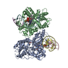 7yc7SC 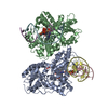 7ycmC  7ycpC  7ycrC 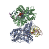 7yd6C 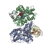 7yd7C 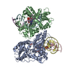 7yd8C 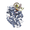 7ydzC 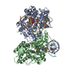 7ye0C 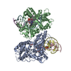 7yebC 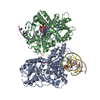 7yecC 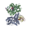 7yeeC 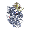 7yeiC 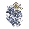 7yejC  7yekC  7yelC  7yemC S: Starting model for refinement C: citing same article ( |
|---|---|
| Similar structure data |
-Links
 PDBj
PDBj


- Assembly
| Deposited unit | 
| ||||||||||
|---|---|---|---|---|---|---|---|---|---|---|---|
| 1 | 
| ||||||||||
| 2 | 
| ||||||||||
| Unit cell |
|
-Components
-Protein , 1 types, 2 molecules AB
| #1: Protein | Photolyase / DNA photolyase Mass: 55123.480 Da / Num. of mol.: 2 / Mutation: M377T Source method: isolated from a genetically manipulated source Source: (gene. exp.) Methanosarcina mazei (archaea)Strain: ATCC BAA-159 / DSM 3647 / Goe1 / Go1 / JCM 11833 / OCM 88 Gene: DU34_19720, DU35_15210, DU36_16165, DU37_08235, DU38_17340, DU39_00605, DU41_17975, DU42_05505, DU44_19145, DU46_16260, DU48_13210, DU49_01325, DU51_15440, DU57_04540, DU59_03190, DU60_01690, ...Gene: DU34_19720, DU35_15210, DU36_16165, DU37_08235, DU38_17340, DU39_00605, DU41_17975, DU42_05505, DU44_19145, DU46_16260, DU48_13210, DU49_01325, DU51_15440, DU57_04540, DU59_03190, DU60_01690, DU61_08490, DU62_04430, DU63_10745, DU65_18915, DU69_04700, DU71_05355, DU72_08110, DU74_09110, FQU78_02295 Production host:  Escherichia coli BL21(DE3) (bacteria) Escherichia coli BL21(DE3) (bacteria)References: UniProt: A0A0F8I5V2, deoxyribodipyrimidine photo-lyase |
|---|
-DNA chain , 2 types, 4 molecules CEDF
| #2: DNA chain | Mass: 4256.767 Da / Num. of mol.: 2 / Source method: obtained synthetically / Source: (synth.) synthetic construct (others) #3: DNA chain | Mass: 4305.805 Da / Num. of mol.: 2 / Source method: obtained synthetically / Source: (synth.) synthetic construct (others) |
|---|
-Non-polymers , 4 types, 568 molecules 


| #4: Chemical | Flavin adenine dinucleotide Mass: 785.550 Da / Num. of mol.: 2 / Source method: obtained synthetically / Formula: C27H33N9O15P2 / Comment: FAD*YM Mass: 785.550 Da / Num. of mol.: 2 / Source method: obtained synthetically / Formula: C27H33N9O15P2 / Comment: FAD*YM#5: Chemical | ChemComp-SO4 / | Sulfate Mass: 96.063 Da / Num. of mol.: 1 / Source method: obtained synthetically / Formula: SO4 Mass: 96.063 Da / Num. of mol.: 1 / Source method: obtained synthetically / Formula: SO4#6: Chemical | ChemComp-GOL / Glycerol Mass: 92.094 Da / Num. of mol.: 9 / Source method: obtained synthetically / Formula: C3H8O3 Mass: 92.094 Da / Num. of mol.: 9 / Source method: obtained synthetically / Formula: C3H8O3#7: Water | ChemComp-HOH / | WaterMass: 18.015 Da / Num. of mol.: 556 / Source method: isolated from a natural source / Formula: H2O |
|---|
-Details
| Has ligand of interest | N |
|---|
-Experimental details
-Experiment
| Experiment | Method: X-RAY DIFFRACTION / Number of used crystals: 1 |
|---|
- Sample preparation
| Crystal | Density Matthews: 2.96 Å3/Da / Density % sol: 58.49 % |
|---|---|
| Crystal grow | Temperature: 296 K / Method: batch mode / pH: 4.6 Details: 100 mM Sodium Acetate pH 4.6 250 mM ammonium sulfate 4% PEG4000 (w/v) 50 mM DTT |
-Data collection
| Diffraction | Mean temperature: 100 K / Serial crystal experiment: N |
|---|---|
| Diffraction source | Source: SYNCHROTRON / Site: SPring-8 / Beamline: BL32XU / Wavelength: 1 Å |
| Detector | Type: RAYONIX MX225HE / Detector: CCD / Date: Jul 30, 2018 |
| Radiation | Protocol: SINGLE WAVELENGTH / Monochromatic (M) / Laue (L): M / Scattering type: x-ray |
| Radiation wavelength | Wavelength: 1 Å / Relative weight: 1 |
| Reflection | Resolution: 2.5→49.95 Å / Num. obs: 48931 / % possible obs: 99.9 % / Redundancy: 26.53 % / Biso Wilson estimate: 3.19 Å2 / CC1/2: 0.739 / Net I/σ(I): 4.66 |
| Reflection shell | Resolution: 2.5→2.65 Å / Num. unique obs: 7737 / CC1/2: 0.437 / % possible all: 100 |
- Processing
| Software |
| ||||||||||||||||||||||||||||||||||||||||||||||||||||||||||||||||||||||||||||||||||||||||||||||||||||||||||||||||||||||||||||||
|---|---|---|---|---|---|---|---|---|---|---|---|---|---|---|---|---|---|---|---|---|---|---|---|---|---|---|---|---|---|---|---|---|---|---|---|---|---|---|---|---|---|---|---|---|---|---|---|---|---|---|---|---|---|---|---|---|---|---|---|---|---|---|---|---|---|---|---|---|---|---|---|---|---|---|---|---|---|---|---|---|---|---|---|---|---|---|---|---|---|---|---|---|---|---|---|---|---|---|---|---|---|---|---|---|---|---|---|---|---|---|---|---|---|---|---|---|---|---|---|---|---|---|---|---|---|---|---|
| Refinement | Method to determine structure: MOLECULAR REPLACEMENT Starting model: 7YC7 Resolution: 2.5→49.29 Å / SU ML: 0.29 / Cross valid method: FREE R-VALUE / σ(F): 1.36 / Phase error: 22.75 / Stereochemistry target values: ML
| ||||||||||||||||||||||||||||||||||||||||||||||||||||||||||||||||||||||||||||||||||||||||||||||||||||||||||||||||||||||||||||||
| Solvent computation | Shrinkage radii: 0.9 Å / VDW probe radii: 1.1 Å / Solvent model: FLAT BULK SOLVENT MODEL | ||||||||||||||||||||||||||||||||||||||||||||||||||||||||||||||||||||||||||||||||||||||||||||||||||||||||||||||||||||||||||||||
| Refinement step | Cycle: LAST / Resolution: 2.5→49.29 Å
| ||||||||||||||||||||||||||||||||||||||||||||||||||||||||||||||||||||||||||||||||||||||||||||||||||||||||||||||||||||||||||||||
| Refine LS restraints |
| ||||||||||||||||||||||||||||||||||||||||||||||||||||||||||||||||||||||||||||||||||||||||||||||||||||||||||||||||||||||||||||||
| LS refinement shell |
|
