 Movie
Movie Controller

[English] 日本語
 Yorodumi
- PDB-8k9f: Cryo-EM structure of the photosynthetic alternative complex III f... -
+ Open data

- Basic information
| Entry | Database: PDB / ID: 8k9f | ||||||
|---|---|---|---|---|---|---|---|
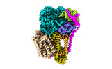
| Title | Cryo-EM structure of the photosynthetic alternative complex III from Chloroflexus aurantiacus at 2.9 angstrom | ||||||
 Components Components |
| ||||||
 Keywords Keywords |  MEMBRANE PROTEIN / Photosynthetic alternative complex III MEMBRANE PROTEIN / Photosynthetic alternative complex III | ||||||
| Function / homology |  Function and homology informationelectron transfer activity / heme binding / membrane / metal ion binding / plasma membrane Function and homology informationelectron transfer activity / heme binding / membrane / metal ion binding / plasma membraneSimilarity search - Function | ||||||
| Biological species |   Chloroflexus aurantiacus (bacteria) Chloroflexus aurantiacus (bacteria) | ||||||
| Method | ELECTRON MICROSCOPY / single particle reconstruction / cryo EM / Resolution: 2.9 Å | ||||||
 Authors Authors | Xu, X. | ||||||
| Funding support |  China, 1items China, 1items
| ||||||
 Citation Citation | Journal: Plant Cell / Year: 2024 Title: Cryo-EM structure of HQNO-bound Alternative Complex III from the anoxygenic phototrophic bacterium Chloroflexus aurantiacus. Authors: Jiyu Xin / Zhenzhen Min / Lu Yu / Xinyi Yuan / Aokun Liu / Wenping Wu / Xin Zhang / Huimin He / Jingyi Wu / Yueyong Xin / Robert E Blankenship / Changlin Tian / Xiaoling Xu /  Abstract: Alternative complex III (ACIII) couples quinol oxidation and electron acceptor reduction with potential transmembrane proton translocation. It is compositionally and structurally different from the ...Alternative complex III (ACIII) couples quinol oxidation and electron acceptor reduction with potential transmembrane proton translocation. It is compositionally and structurally different from the cytochrome bc1/b6f complexes, but functionally replaces these enzymes in the photosynthetic and/or respiratory electron transport chains (ETCs) of many bacteria. However, the true compositions and architectures of ACIIIs remain unclear, as do their structural and functional relevance in mediating the ETCs. We here determined cryogenic electron microscopy structures of photosynthetic ACIII isolated from Chloroflexus aurantiacus (CaACIIIp), in apo-form and in complexed form bound to a menadiol analog 2-heptyl-4-hydroxyquinoline-N-oxide (HQNO). Besides six canonical subunits (ActABCDEF), the structures revealed conformations of two previously unresolved subunits, ActG and I, which contributed to the complex stability. We also elucidated the structural basis of menaquinol oxidation and subsequent electron transfer along the [3Fe-4S]-6 hemes wire to its periplasmic electron acceptors, using electron paramagnetic resonance (EPR), spectroelectrochemistry, enzymatic analyses and molecular dynamics (MD) simulations. A unique insertion loop in ActE was shown to function in determining the binding specificity of CaACIIIp for downstream electron acceptors. This study broadens our understanding of the structural diversity and molecular evolution of ACIIIs, enabling further investigation of the (mena)quinol oxidoreductases evolved coupling mechanism in bacterial energy conservation. | ||||||
| History |
|
- Structure visualization
| Structure viewer | Molecule: MolmilJmol/JSmol |
|---|
- Downloads & links
-Download
| PDBx/mmCIF format | 8k9f.cif.gz | 513.2 KB | Display | PDBx/mmCIF format |
|---|---|---|---|---|
| PDB format | pdb8k9f.ent.gz | 405 KB | Display | PDB format |
| PDBx/mmJSON format | 8k9f.json.gz | Tree view | PDBx/mmJSON format | |
| Others |  Other downloads Other downloads |
-Validation report
| Arichive directory | https://data.pdbj.org/pub/pdb/validation_reports/k9/8k9fftp://data.pdbj.org/pub/pdb/validation_reports/k9/8k9f | HTTPS FTP |
|---|
-Related structure data
| Related structure data | 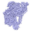 36985MC 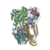 8k9eC 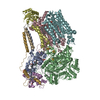 8x2jC M: map data used to model this data C: citing same article ( |
|---|---|
| Similar structure data |
-Links
 PDBj
PDBj


- Assembly
| Deposited unit | 
|
|---|---|
| 1 |
|
-Components
-Protein , 5 types, 5 molecules ABCEG
| #1: Protein | Mass: 25247.039 Da / Num. of mol.: 1 / Source method: isolated from a natural source Source: (natural) Chloroflexus aurantiacus (strain ATCC 29366 / DSM 635 / J-10-fl) (bacteria)References: UniProt: A9WEV2 |
|---|---|
| #2: Protein | Mass: 113115.359 Da / Num. of mol.: 1 / Source method: isolated from a natural source Source: (natural) Chloroflexus aurantiacus (strain ATCC 29366 / DSM 635 / J-10-fl) (bacteria)References: UniProt: A9WEV3 |
| #3: Protein | Mass: 55223.520 Da / Num. of mol.: 1 / Source method: isolated from a natural source Source: (natural) Chloroflexus aurantiacus (strain ATCC 29366 / DSM 635 / J-10-fl) (bacteria)References: UniProt: A9WEV4 |
| #5: Protein | Mass: 23017.016 Da / Num. of mol.: 1 / Source method: isolated from a natural source Source: (natural) Chloroflexus aurantiacus (strain ATCC 29366 / DSM 635 / J-10-fl) (bacteria)References: UniProt: A9WEV6 |
| #7: Protein | Mass: 12485.551 Da / Num. of mol.: 1 / Source method: isolated from a natural source Source: (natural) Chloroflexus aurantiacus (strain ATCC 29366 / DSM 635 / J-10-fl) (bacteria)References: UniProt: A9WEV8 |
-Quinol:cytochrome c oxidoreductase ... , 2 types, 2 molecules DF
| #4: Protein | Mass: 19648.570 Da / Num. of mol.: 1 / Source method: isolated from a natural source Source: (natural) Chloroflexus aurantiacus (strain ATCC 29366 / DSM 635 / J-10-fl) (bacteria)References: UniProt: A9WEV5 |
|---|---|
| #6: Protein | Mass: 45721.672 Da / Num. of mol.: 1 / Source method: isolated from a natural source Source: (natural) Chloroflexus aurantiacus (strain ATCC 29366 / DSM 635 / J-10-fl) (bacteria)References: UniProt: A9WEV7 |
-Protein/peptide , 1 types, 1 molecules I
| #8: Protein/peptide | Mass: 4322.134 Da / Num. of mol.: 1 / Source method: isolated from a natural source Source: (natural) Chloroflexus aurantiacus (strain ATCC 29366 / DSM 635 / J-10-fl) (bacteria) |
|---|
-Non-polymers , 7 types, 14 molecules 


| #9: Chemical | ChemComp-HEC / Heme C Mass: 618.503 Da / Num. of mol.: 6 / Source method: obtained synthetically / Formula: C34H34FeN4O4 Mass: 618.503 Da / Num. of mol.: 6 / Source method: obtained synthetically / Formula: C34H34FeN4O4#10: Chemical | Iron–sulfur cluster Mass: 351.640 Da / Num. of mol.: 3 / Source method: obtained synthetically / Formula: Fe4S4 Mass: 351.640 Da / Num. of mol.: 3 / Source method: obtained synthetically / Formula: Fe4S4#11: Chemical | ChemComp-F3S / | Iron–sulfur cluster Mass: 295.795 Da / Num. of mol.: 1 / Source method: obtained synthetically / Formula: Fe3S4 Mass: 295.795 Da / Num. of mol.: 1 / Source method: obtained synthetically / Formula: Fe3S4#12: Chemical | ChemComp-MG / |  Mass: 24.305 Da / Num. of mol.: 1 / Source method: obtained synthetically / Formula: Mg Mass: 24.305 Da / Num. of mol.: 1 / Source method: obtained synthetically / Formula: Mg#13: Chemical | ChemComp-JLQ / [( | Mass: 663.906 Da / Num. of mol.: 1 / Source method: obtained synthetically / Formula: C35H70NO8P #14: Chemical | ChemComp-JL3 / [( | Mass: 663.906 Da / Num. of mol.: 1 / Source method: obtained synthetically / Formula: C35H70NO8P #15: Chemical | ChemComp-JM9 / | Mass: 765.240 Da / Num. of mol.: 1 / Source method: obtained synthetically / Formula: C48H92O6 |
|---|
-Details
| Has ligand of interest | N |
|---|
-Experimental details
-Experiment
| Experiment | Method: ELECTRON MICROSCOPY |
|---|---|
| EM experiment | Aggregation state: PARTICLE / 3D reconstruction method: single particle reconstruction |
- Sample preparation
| Component | Name: Alternative complex III / Type: COMPLEX / Entity ID: #1-#8 / Source: NATURAL |
|---|---|
| Source (natural) | Organism: Chloroflexus aurantiacus (strain ATCC 29366 / DSM 635 / J-10-fl) (bacteria) |
| Buffer solution | pH: 8 |
| Specimen | Embedding applied: NO / Shadowing applied: NO / Staining applied: NO / Vitrification applied: YES |
| Vitrification | Cryogen name: ETHANE |
- Electron microscopy imaging
| Experimental equipment |  Model: Titan Krios / Image courtesy: FEI Company |
|---|---|
| Microscopy | Model: FEI TITAN KRIOS |
| Electron gun | Electron source: FIELD EMISSION GUN / Accelerating voltage: 300 kV / Illumination mode: FLOOD BEAM |
| Electron lens | Mode: BRIGHT FIELDBright-field microscopy / Nominal defocus max: 1800 nm / Nominal defocus min: 1000 nm |
| Image recording | Electron dose: 50 e/Å2 / Film or detector model: FEI FALCON IV (4k x 4k) |
- Processing
| CTF correction | Type: PHASE FLIPPING AND AMPLITUDE CORRECTION | ||||||||||||||||||||||||
|---|---|---|---|---|---|---|---|---|---|---|---|---|---|---|---|---|---|---|---|---|---|---|---|---|---|
| 3D reconstruction | Resolution: 2.9 Å / Resolution method: FSC 0.143 CUT-OFF / Num. of particles: 103633 / Symmetry type: POINT | ||||||||||||||||||||||||
| Refine LS restraints |
|
