 Movie
Movie Controller


+ Open data

- Basic information
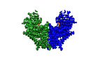
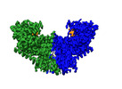
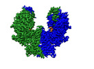
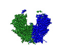
| Entry | Database: PDB / ID: 8k3w | ||||||
|---|---|---|---|---|---|---|---|
| Title | S. cerevisiae Chs1 in complex with UDP-GlcNAc and GlcNAc | ||||||
 Components Components | Chitin synthase 1 | ||||||
 Keywords Keywords | ANTIFUNGAL PROTEIN/INHIBITOR / ANTIFUNGAL PROTEIN-INHIBITOR complex | ||||||
| Function / homology |  Function and homology information Function and homology informationchitosome / cell wall chitin biosynthetic process / chitin synthase / chitin synthase activity / septum digestion after cytokinesis / cell septum / cell periphery / cell wall organization / plasma membraneSimilarity search - Function | ||||||
| Biological species |  Saccharomyces cerevisiae (brewer's yeast) Saccharomyces cerevisiae (brewer's yeast) | ||||||
| Method | ELECTRON MICROSCOPY / single particle reconstruction / cryo EM / Resolution: 2.91 Å | ||||||
 Authors Authors | Bai, L. / Chen, D. | ||||||
| Funding support |  China, 1items China, 1items
| ||||||
 Citation Citation | Journal: Nat Commun / Year: 2023 Title: Structure, catalysis, chitin transport, and selective inhibition of chitin synthase. Authors: Dan-Dan Chen / Zhao-Bin Wang / Le-Xuan Wang / Peng Zhao / Cai-Hong Yun / Lin Bai / Abstract: Chitin is one of the most abundant natural biopolymers and serves as a critical structural component of extracellular matrices, including fungal cell walls and insect exoskeletons. As a linear ...Chitin is one of the most abundant natural biopolymers and serves as a critical structural component of extracellular matrices, including fungal cell walls and insect exoskeletons. As a linear polymer of β-(1,4)-linked N-acetylglucosamine, chitin is synthesized by chitin synthases, which are recognized as targets for antifungal and anti-insect drugs. In this study, we determine seven different cryo-electron microscopy structures of a Saccharomyces cerevisiae chitin synthase in the absence and presence of glycosyl donor, acceptor, product, or peptidyl nucleoside inhibitors. Combined with functional analyses, these structures show how the donor and acceptor substrates bind in the active site, how substrate hydrolysis drives self-priming, how a chitin-conducting transmembrane channel opens, and how peptidyl nucleoside inhibitors inhibit chitin synthase. Our work provides a structural basis for understanding the function and inhibition of chitin synthase. | ||||||
| History |
|
- Structure visualization
| Structure viewer | Molecule: MolmilJmol/JSmol |
|---|
- Downloads & links
-Download
| PDBx/mmCIF format | 8k3w.cif.gz | 270.6 KB | Display | PDBx/mmCIF format |
|---|---|---|---|---|
| PDB format | pdb8k3w.ent.gz | 203.4 KB | Display | PDB format |
| PDBx/mmJSON format | 8k3w.json.gz | Tree view | PDBx/mmJSON format | |
| Others |  Other downloads Other downloads |
-Validation report
| Arichive directory | https://data.pdbj.org/pub/pdb/validation_reports/k3/8k3wftp://data.pdbj.org/pub/pdb/validation_reports/k3/8k3w | HTTPS FTP |
|---|
-Related structure data
| Related structure data | 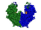 36863MC  8k3pC  8k3qC  8k3rC  8k3tC  8k3uC  8k3vC 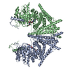 8k3xC M: map data used to model this data C: citing same article ( |
|---|---|
| Similar structure data |
-Links
 PDBj
PDBj

- Assembly
| Deposited unit | 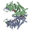
|
|---|---|
| 1 |
|
-Components
| #1: Protein | / Chitin-UDP acetyl-glucosaminyl transferase 1 Mass: 130001.141 Da / Num. of mol.: 2 Source method: isolated from a genetically manipulated source Source: (gene. exp.) Saccharomyces cerevisiae (brewer's yeast)Gene: CHS1 / Production host: Saccharomyces cerevisiae (brewer's yeast) / References: UniProt: P08004, chitin synthase#2: Chemical |   Mass: 24.305 Da / Num. of mol.: 2 / Source method: obtained synthetically / Formula: Mg / Feature type: SUBJECT OF INVESTIGATION Mass: 24.305 Da / Num. of mol.: 2 / Source method: obtained synthetically / Formula: Mg / Feature type: SUBJECT OF INVESTIGATION#3: Chemical | POPC  Mass: 760.076 Da / Num. of mol.: 2 / Source method: obtained synthetically / Formula: C42H82NO8P / Feature type: SUBJECT OF INVESTIGATION / Comment: phospholipid*YM Mass: 760.076 Da / Num. of mol.: 2 / Source method: obtained synthetically / Formula: C42H82NO8P / Feature type: SUBJECT OF INVESTIGATION / Comment: phospholipid*YM#4: Chemical |   Mass: 607.354 Da / Num. of mol.: 2 / Source method: obtained synthetically / Formula: C17H27N3O17P2 / Feature type: SUBJECT OF INVESTIGATION Mass: 607.354 Da / Num. of mol.: 2 / Source method: obtained synthetically / Formula: C17H27N3O17P2 / Feature type: SUBJECT OF INVESTIGATIONHas ligand of interest | Y | |
|---|
-Experimental details
-Experiment
| Experiment | Method: ELECTRON MICROSCOPY |
|---|---|
| EM experiment | Aggregation state: PARTICLE / 3D reconstruction method: single particle reconstruction |
- Sample preparation
| Component | Name: S. cerevisiae Chs1 in complex with UDP-GlcNAc and GlcNAc Type: COMPLEX / Entity ID: #1 / Source: RECOMBINANT |
|---|---|
| Source (natural) | Organism: Saccharomyces cerevisiae (brewer's yeast) |
| Source (recombinant) | Organism: Saccharomyces cerevisiae (brewer's yeast) |
| Buffer solution | pH: 7.4 |
| Specimen | Conc.: 4 mg/ml / Embedding applied: NO / Shadowing applied: NO / Staining applied: NO / Vitrification applied: YES |
| Vitrification | Cryogen name: ETHANE |
- Electron microscopy imaging
| Experimental equipment |  Model: Titan Krios / Image courtesy: FEI Company |
|---|---|
| Microscopy | Model: FEI TITAN KRIOS |
| Electron gun | Electron source: FIELD EMISSION GUN / Accelerating voltage: 300 kV / Illumination mode: OTHER |
| Electron lens | Mode: BRIGHT FIELDBright-field microscopy / Nominal defocus max: 1800 nm / Nominal defocus min: 1200 nm |
| Image recording | Electron dose: 50 e/Å2 / Film or detector model: GATAN K3 (6k x 4k) |
- Processing
| EM software | Name: PHENIX / Version: 1.18.2_3874: / Category: model refinement | ||||||||||||||||||||||||
|---|---|---|---|---|---|---|---|---|---|---|---|---|---|---|---|---|---|---|---|---|---|---|---|---|---|
| CTF correction | Type: NONE | ||||||||||||||||||||||||
| 3D reconstruction | Resolution: 2.91 Å / Resolution method: FSC 0.143 CUT-OFF / Num. of particles: 107259 / Symmetry type: POINT | ||||||||||||||||||||||||
| Refine LS restraints |
|
