 Movie
Movie Controller

[English] 日本語
 Yorodumi
- PDB-8jv8: Cryo-EM structure of the panda P2X7 receptor in complex with PPNDS -
+ Open data

- Basic information
| Entry | Database: PDB / ID: 8jv8 | ||||||
|---|---|---|---|---|---|---|---|
| Title | Cryo-EM structure of the panda P2X7 receptor in complex with PPNDS | ||||||
 Components Components | P2X purinoceptor | ||||||
 Keywords Keywords |  TRANSPORT PROTEIN / Channel TRANSPORT PROTEIN / Channel | ||||||
| Function / homology |  Function and homology information Function and homology informationpurinergic nucleotide receptor activity / extracellularly ATP-gated monoatomic cation channel activity / response to ATP / calcium ion transmembrane transport / postsynapse / ATP binding / plasma membraneSimilarity search - Function | ||||||
| Biological species |  Ailuropoda melanoleuca (giant panda) Ailuropoda melanoleuca (giant panda) | ||||||
| Method | ELECTRON MICROSCOPY / single particle reconstruction / cryo EM / Resolution: 3.34 Å | ||||||
 Authors Authors | Sheng, D. / Hattori, M. | ||||||
| Funding support |  China, 1items China, 1items
| ||||||
 Citation Citation | Journal: Elife / Year: 2024 Title: Structural insights into the orthosteric inhibition of P2X receptors by non-ATP analog antagonists. Authors: Danqi Sheng / Chen-Xi Yue / Fei Jin / Yao Wang / Muneyoshi Ichikawa / Ye Yu / Chang-Run Guo / Motoyuki Hattori / Abstract: P2X receptors are extracellular ATP-gated ion channels that form homo- or heterotrimers and consist of seven subtypes. They are expressed in various tissues, including neuronal and nonneuronal cells, ...P2X receptors are extracellular ATP-gated ion channels that form homo- or heterotrimers and consist of seven subtypes. They are expressed in various tissues, including neuronal and nonneuronal cells, and play critical roles in physiological processes such as neurotransmission, inflammation, pain, and cancer. As a result, P2X receptors have attracted considerable interest as drug targets, and various competitive inhibitors have been developed. However, although several P2X receptor structures from different subtypes have been reported, the limited structural information of P2X receptors in complex with competitive antagonists hampers the understanding of orthosteric inhibition, hindering the further design and optimization of those antagonists for drug discovery. We determined the cryogenic electron microscopy (cryo-EM) structures of the mammalian P2X7 receptor in complex with two classical competitive antagonists of pyridoxal-5'-phosphate derivatives, pyridoxal-5'-phosphate-6-(2'-naphthylazo-6'-nitro-4',8'-disulfonate) (PPNDS) and pyridoxal phosphate-6-azophenyl-2',5'-disulfonic acid (PPADS), and performed structure-based mutational analysis by patch-clamp recording as well as molecular dynamics (MD) simulations. Our structures revealed the orthosteric site for PPADS/PPNDS, and structural comparison with the previously reported apo- and ATP-bound structures showed how PPADS/PPNDS binding inhibits the conformational changes associated with channel activation. In addition, structure-based mutational analysis identified key residues involved in the PPNDS sensitivity of P2X1 and P2X3, which are known to have higher affinity for PPADS/PPNDS than other P2X subtypes. #1: Journal: Elife / Year: 2023Title: Structural insights into the orthosteric inhibition of P2X receptors by non-ATP-analog antagonists Authors: Sheng, D. / Yue, C. / Jin, F. / Wang, Y. / Ichikawa, M. / Yu, Y. / Guo, C.R. / Hattori, M. | ||||||
| History |
|
- Structure visualization
| Structure viewer | Molecule: MolmilJmol/JSmol |
|---|
- Downloads & links
-Download
| PDBx/mmCIF format | 8jv8.cif.gz | 197.7 KB | Display | PDBx/mmCIF format |
|---|---|---|---|---|
| PDB format | pdb8jv8.ent.gz | 153.1 KB | Display | PDB format |
| PDBx/mmJSON format | 8jv8.json.gz | Tree view | PDBx/mmJSON format | |
| Others |  Other downloads Other downloads |
-Validation report
| Arichive directory | https://data.pdbj.org/pub/pdb/validation_reports/jv/8jv8ftp://data.pdbj.org/pub/pdb/validation_reports/jv/8jv8 | HTTPS FTP |
|---|
-Related structure data
| Related structure data | 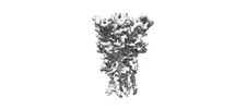 36671MC 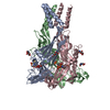 8jv7C M: map data used to model this data C: citing same article ( |
|---|---|
| Similar structure data |

-Links
 PDBj
PDBj- Assembly
| Deposited unit | 
|
|---|---|
| 1 |
|
-Components
| #1: Protein | Mass: 38716.234 Da / Num. of mol.: 3 / Mutation: V35A, R125A, E174K, N241S, N284S Source method: isolated from a genetically manipulated source Source: (gene. exp.) Ailuropoda melanoleuca (giant panda) / Gene: PANDA_000894 / Production host:   Spodoptera frugiperda (fall armyworm) / References: UniProt: D2GVW0 Spodoptera frugiperda (fall armyworm) / References: UniProt: D2GVW0#2: Sugar | ChemComp-NAG / N-Acetylglucosamine  Type: D-saccharide, beta linking / Mass: 221.208 Da / Num. of mol.: 6 Type: D-saccharide, beta linking / Mass: 221.208 Da / Num. of mol.: 6Source method: isolated from a genetically manipulated source Formula: C8H15NO6 #3: Chemical |   Mass: 606.434 Da / Num. of mol.: 3 / Source method: obtained synthetically / Formula: C18H15N4O14PS2 / Feature type: SUBJECT OF INVESTIGATION Mass: 606.434 Da / Num. of mol.: 3 / Source method: obtained synthetically / Formula: C18H15N4O14PS2 / Feature type: SUBJECT OF INVESTIGATION#4: Water | ChemComp-HOH / | Water Mass: 18.015 Da / Num. of mol.: 3 / Source method: isolated from a natural source / Formula: H2O Mass: 18.015 Da / Num. of mol.: 3 / Source method: isolated from a natural source / Formula: H2OHas ligand of interest | Y | |
|---|
-Experimental details
-Experiment
| Experiment | Method: ELECTRON MICROSCOPY |
|---|---|
| EM experiment | Aggregation state: PARTICLE / 3D reconstruction method: single particle reconstruction |
- Sample preparation
| Component | Name: P2X7 trimer / Type: COMPLEX / Entity ID: #1 / Source: RECOMBINANT |
|---|---|
| Molecular weight | Experimental value: NO |
| Source (natural) | Organism: Ailuropoda melanoleuca (giant panda) |
| Source (recombinant) | Organism: Spodoptera frugiperda (fall armyworm) |
| Buffer solution | pH: 7.5 |
| Specimen | Embedding applied: NO / Shadowing applied: NO / Staining applied: NO / Vitrification applied: YES |
| Vitrification | Cryogen name: ETHANE |
- Electron microscopy imaging
| Experimental equipment |  Model: Titan Krios / Image courtesy: FEI Company |
|---|---|
| Microscopy | Model: FEI TITAN KRIOS |
| Electron gun | Electron source: FIELD EMISSION GUN / Accelerating voltage: 300 kV / Illumination mode: FLOOD BEAM |
| Electron lens | Mode: BRIGHT FIELDBright-field microscopy / Nominal defocus max: 2000 nm / Nominal defocus min: 1300 nm |
| Image recording | Electron dose: 50 e/Å2 / Film or detector model: GATAN K3 (6k x 4k) |
- Processing
| CTF correction | Type: PHASE FLIPPING AND AMPLITUDE CORRECTION | ||||||||||||||||||||||||
|---|---|---|---|---|---|---|---|---|---|---|---|---|---|---|---|---|---|---|---|---|---|---|---|---|---|
| 3D reconstruction | Resolution: 3.34 Å / Resolution method: FSC 0.143 CUT-OFF / Num. of particles: 121008 / Symmetry type: POINT | ||||||||||||||||||||||||
| Refine LS restraints |
|
