 Movie
Movie Controller

[English] 日本語
 Yorodumi
- PDB-8jrv: Cryo-EM structure of the glucagon receptor bound to glucagon and ... -
+ Open data

- Basic information
| Entry | Database: PDB / ID: 8jrv | |||||||||
|---|---|---|---|---|---|---|---|---|---|---|
| Title | Cryo-EM structure of the glucagon receptor bound to glucagon and beta-arrestin 1 | |||||||||
 Components Components |
| |||||||||
 Keywords Keywords |  MEMBRANE PROTEIN / Complex structure / glucagon receptor / beta-arrestin 1 / glucagon MEMBRANE PROTEIN / Complex structure / glucagon receptor / beta-arrestin 1 / glucagon | |||||||||
| Function / homology |  Function and homology informationprotein C (activated) / renal water retention / Defective AVP does not bind AVPR2 and causes neurohypophyseal diabetes insipidus (NDI) / positive regulation of establishment of endothelial barrier / Vasopressin-like receptors / regulation of systemic arterial blood pressure by vasopressin / vasopressin receptor activity / glucagon receptor binding / negative regulation of coagulation / : ...protein C (activated) / renal water retention / Defective AVP does not bind AVPR2 and causes neurohypophyseal diabetes insipidus (NDI) / positive regulation of establishment of endothelial barrier / Vasopressin-like receptors / regulation of systemic arterial blood pressure by vasopressin / vasopressin receptor activity / glucagon receptor binding / negative regulation of coagulation / : / regulation of glycogen metabolic process / glucagon receptor activity / hemostasis / positive regulation of systemic arterial blood pressure / telencephalon development / negative regulation of execution phase of apoptosis / negative regulation of blood coagulation / feeding behavior / positive regulation of intracellular signal transduction / response to starvation / positive regulation of calcium ion import / cellular response to glucagon stimulus / exocytosis / positive regulation of insulin secretion involved in cellular response to glucose stimulus / peptide hormone binding / regulation of insulin secretion / endocytic vesicle / Synthesis, secretion, and deacylation of Ghrelin / Gamma-carboxylation of protein precursors / Transport of gamma-carboxylated protein precursors from the endoplasmic reticulum to the Golgi apparatus / Common Pathway of Fibrin Clot Formation / Removal of aminoterminal propeptides from gamma-carboxylated proteins / positive regulation of gluconeogenesis / positive regulation of vasoconstriction / cellular response to hormone stimulus / protein kinase A signaling / activation of adenylate cyclase activity / Intrinsic Pathway of Fibrin Clot Formation / cellular response to starvation / hormone-mediated signaling pathway / response to nutrient / guanyl-nucleotide exchange factor activity / viral budding from plasma membrane / response to activity / positive regulation of peptidyl-threonine phosphorylation / generation of precursor metabolites and energy / response to cytokine / gluconeogenesis / Cell surface interactions at the vascular wall / peptide binding / Post-translational protein phosphorylation / clathrin-coated endocytic vesicle membrane / adenylate cyclase-modulating G protein-coupled receptor signaling pathway / Glucagon signaling in metabolic regulation / adenylate cyclase-activating G protein-coupled receptor signaling pathway / hormone activity / Synthesis, secretion, and inactivation of Glucagon-like Peptide-1 (GLP-1) / Glucagon-type ligand receptors / regulation of blood pressure / Vasopressin regulates renal water homeostasis via Aquaporins / negative regulation of inflammatory response / Golgi lumen / Glucagon-like Peptide-1 (GLP1) regulates insulin secretion / Regulation of Insulin-like Growth Factor (IGF) transport and uptake by Insulin-like Growth Factor Binding Proteins (IGFBPs) / blood coagulation / Cargo recognition for clathrin-mediated endocytosis / positive regulation of peptidyl-serine phosphorylation / glucose homeostasis / Clathrin-mediated endocytosis / G alpha (s) signalling events / G alpha (q) signalling events / clathrin-dependent endocytosis of virus by host cell / secretory granule lumen / cell surface receptor signaling pathway / positive regulation of ERK1 and ERK2 cascade / host cell surface receptor binding / endosome / G protein-coupled receptor signaling pathway / negative regulation of cell population proliferation / fusion of virus membrane with host plasma membrane / endoplasmic reticulum lumen / signaling receptor binding / serine-type endopeptidase activity / fusion of virus membrane with host endosome membrane / viral envelope / calcium ion binding / positive regulation of cell population proliferation / virion attachment to host cell / positive regulation of gene expression / negative regulation of apoptotic process / host cell plasma membrane / virion membrane / perinuclear region of cytoplasm / Golgi apparatus / endoplasmic reticulum / proteolysis / extracellular space / extracellular region / membrane / identical protein binding Function and homology informationprotein C (activated) / renal water retention / Defective AVP does not bind AVPR2 and causes neurohypophyseal diabetes insipidus (NDI) / positive regulation of establishment of endothelial barrier / Vasopressin-like receptors / regulation of systemic arterial blood pressure by vasopressin / vasopressin receptor activity / glucagon receptor binding / negative regulation of coagulation / : ...protein C (activated) / renal water retention / Defective AVP does not bind AVPR2 and causes neurohypophyseal diabetes insipidus (NDI) / positive regulation of establishment of endothelial barrier / Vasopressin-like receptors / regulation of systemic arterial blood pressure by vasopressin / vasopressin receptor activity / glucagon receptor binding / negative regulation of coagulation / : / regulation of glycogen metabolic process / glucagon receptor activity / hemostasis / positive regulation of systemic arterial blood pressure / telencephalon development / negative regulation of execution phase of apoptosis / negative regulation of blood coagulation / feeding behavior / positive regulation of intracellular signal transduction / response to starvation / positive regulation of calcium ion import / cellular response to glucagon stimulus / exocytosis / positive regulation of insulin secretion involved in cellular response to glucose stimulus / peptide hormone binding / regulation of insulin secretion / endocytic vesicle / Synthesis, secretion, and deacylation of Ghrelin / Gamma-carboxylation of protein precursors / Transport of gamma-carboxylated protein precursors from the endoplasmic reticulum to the Golgi apparatus / Common Pathway of Fibrin Clot Formation / Removal of aminoterminal propeptides from gamma-carboxylated proteins / positive regulation of gluconeogenesis / positive regulation of vasoconstriction / cellular response to hormone stimulus / protein kinase A signaling / activation of adenylate cyclase activity / Intrinsic Pathway of Fibrin Clot Formation / cellular response to starvation / hormone-mediated signaling pathway / response to nutrient / guanyl-nucleotide exchange factor activity / viral budding from plasma membrane / response to activity / positive regulation of peptidyl-threonine phosphorylation / generation of precursor metabolites and energy / response to cytokine / gluconeogenesis / Cell surface interactions at the vascular wall / peptide binding / Post-translational protein phosphorylation / clathrin-coated endocytic vesicle membrane / adenylate cyclase-modulating G protein-coupled receptor signaling pathway / Glucagon signaling in metabolic regulation / adenylate cyclase-activating G protein-coupled receptor signaling pathway / hormone activity / Synthesis, secretion, and inactivation of Glucagon-like Peptide-1 (GLP-1) / Glucagon-type ligand receptors / regulation of blood pressure / Vasopressin regulates renal water homeostasis via Aquaporins / negative regulation of inflammatory response / Golgi lumen / Glucagon-like Peptide-1 (GLP1) regulates insulin secretion / Regulation of Insulin-like Growth Factor (IGF) transport and uptake by Insulin-like Growth Factor Binding Proteins (IGFBPs) / blood coagulation / Cargo recognition for clathrin-mediated endocytosis / positive regulation of peptidyl-serine phosphorylation / glucose homeostasis / Clathrin-mediated endocytosis / G alpha (s) signalling events / G alpha (q) signalling events / clathrin-dependent endocytosis of virus by host cell / secretory granule lumen / cell surface receptor signaling pathway / positive regulation of ERK1 and ERK2 cascade / host cell surface receptor binding / endosome / G protein-coupled receptor signaling pathway / negative regulation of cell population proliferation / fusion of virus membrane with host plasma membrane / endoplasmic reticulum lumen / signaling receptor binding / serine-type endopeptidase activity / fusion of virus membrane with host endosome membrane / viral envelope / calcium ion binding / positive regulation of cell population proliferation / virion attachment to host cell / positive regulation of gene expression / negative regulation of apoptotic process / host cell plasma membrane / virion membrane / perinuclear region of cytoplasm / Golgi apparatus / endoplasmic reticulum / proteolysis / extracellular space / extracellular region / membrane / identical protein bindingSimilarity search - Function | |||||||||
| Biological species |   Influenza A virus Influenza A virus Homo sapiens (human)Escherichia phage EcSzw-2 (virus) Homo sapiens (human)Escherichia phage EcSzw-2 (virus) | |||||||||
| Method | ELECTRON MICROSCOPY / single particle reconstruction / cryo EM / Resolution: 3.3 Å | |||||||||
 Authors Authors | Chen, K. / Zhang, C. / Lin, S. / Zhao, Q. / Wu, B. | |||||||||
| Funding support |  China, 2items China, 2items
| |||||||||
 Citation Citation | Journal: Nature / Year: 2023 Title: Tail engagement of arrestin at the glucagon receptor. Authors: Kun Chen / Chenhui Zhang / Shuling Lin / Xinyu Yan / Heng Cai / Cuiying Yi / Limin Ma / Xiaojing Chu / Yuchen Liu / Ya Zhu / Shuo Han / Qiang Zhao / Beili Wu / Abstract: Arrestins have pivotal roles in regulating G protein-coupled receptor (GPCR) signalling by desensitizing G protein activation and mediating receptor internalization. It has been proposed that the ...Arrestins have pivotal roles in regulating G protein-coupled receptor (GPCR) signalling by desensitizing G protein activation and mediating receptor internalization. It has been proposed that the arrestin binds to the receptor in two different conformations, 'tail' and 'core', which were suggested to govern distinct processes of receptor signalling and trafficking. However, little structural information is available for the tail engagement of the arrestins. Here we report two structures of the glucagon receptor (GCGR) bound to β-arrestin 1 (βarr1) in glucagon-bound and ligand-free states. These structures reveal a receptor tail-engaged binding mode of βarr1 with many unique features, to our knowledge, not previously observed. Helix VIII, instead of the receptor core, has a major role in accommodating βarr1 by forming extensive interactions with the central crest of βarr1. The tail-binding pose is further defined by a close proximity between the βarr1 C-edge and the receptor helical bundle, and stabilized by a phosphoinositide derivative that bridges βarr1 with helices I and VIII of GCGR. Lacking any contact with the arrestin, the receptor core is in an inactive state and loosely binds to glucagon. Further functional studies suggest that the tail conformation of GCGR-βarr governs βarr recruitment at the plasma membrane and endocytosis of GCGR, and provides a molecular basis for the receptor forming a super-complex simultaneously with G protein and βarr to promote sustained signalling within endosomes. These findings extend our knowledge about the arrestin-mediated modulation of GPCR functionalities. | |||||||||
| History |
|
- Structure visualization
| Structure viewer | Molecule: MolmilJmol/JSmol |
|---|
- Downloads & links
-Download
| PDBx/mmCIF format | 8jrv.cif.gz | 248.2 KB | Display | PDBx/mmCIF format |
|---|---|---|---|---|
| PDB format | pdb8jrv.ent.gz | 170 KB | Display | PDB format |
| PDBx/mmJSON format | 8jrv.json.gz | Tree view | PDBx/mmJSON format | |
| Others |  Other downloads Other downloads |
-Validation report
| Arichive directory | https://data.pdbj.org/pub/pdb/validation_reports/jr/8jrvftp://data.pdbj.org/pub/pdb/validation_reports/jr/8jrv | HTTPS FTP |
|---|
-Related structure data
| Related structure data | 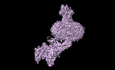 36607MC 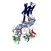 8jruC M: map data used to model this data C: citing same article ( |
|---|---|
| Similar structure data |

-Links
 PDBj
PDBj


- Assembly
| Deposited unit | 
|
|---|---|
| 1 |
|
-Components
| #1: Protein | Mass: 54163.012 Da / Num. of mol.: 1 Source method: isolated from a genetically manipulated source Source: (gene. exp.) Influenza A virus (strain A/Victoria/3/1975 H3N2), (gene. exp.) Homo sapiens (human)Gene: HA, PROC, GCGR, AVPR2, ADHR, DIR, DIR3, V2R / Production host:   Spodoptera frugiperda (fall armyworm) Spodoptera frugiperda (fall armyworm)References: UniProt: P03435, UniProt: P04070, UniProt: P47871, UniProt: P30518 | ||||||
|---|---|---|---|---|---|---|---|
| #2: Protein/peptide | Mass: 3486.781 Da / Num. of mol.: 1 / Source method: obtained synthetically / Source: (synth.) Homo sapiens (human) / References: UniProt: P01275 | ||||||
| #3: Antibody | Mass: 69173.891 Da / Num. of mol.: 3 Source method: isolated from a genetically manipulated source Source: (gene. exp.) Homo sapiens (human) / Production host: Spodoptera frugiperda (fall armyworm)#4: Antibody | | Single-domain antibodyMass: 13867.408 Da / Num. of mol.: 1 Source method: isolated from a genetically manipulated source Source: (gene. exp.) Escherichia phage EcSzw-2 (virus) / Production host:  Escherichia coli BL21 (bacteria) Escherichia coli BL21 (bacteria)#5: Chemical | ChemComp-PIO / [( |   Mass: 746.566 Da / Num. of mol.: 1 / Source method: obtained synthetically / Formula: C25H49O19P3 / Feature type: SUBJECT OF INVESTIGATION Mass: 746.566 Da / Num. of mol.: 1 / Source method: obtained synthetically / Formula: C25H49O19P3 / Feature type: SUBJECT OF INVESTIGATIONHas ligand of interest | Y | |
-Experimental details
-Experiment
| Experiment | Method: ELECTRON MICROSCOPY |
|---|---|
| EM experiment | Aggregation state: PARTICLE / 3D reconstruction method: single particle reconstruction |
- Sample preparation
| Component | Name: The glucagon receptor bound to glucagon and beta-arrestin 1 Type: COMPLEX / Entity ID: #1-#4 / Source: MULTIPLE SOURCES |
|---|---|
| Source (natural) | Organism: Homo sapiens (human) |
| Source (recombinant) | Organism: Spodoptera frugiperda (fall armyworm) |
| Buffer solution | pH: 7.5 |
| Specimen | Conc.: 6 mg/ml / Embedding applied: NO / Shadowing applied: NO / Staining applied: NO / Vitrification applied: YES |
| Vitrification | Cryogen name: ETHANE |
- Electron microscopy imaging
| Experimental equipment |  Model: Titan Krios / Image courtesy: FEI Company |
|---|---|
| Microscopy | Model: FEI TITAN KRIOS |
| Electron gun | Electron source: FIELD EMISSION GUN / Accelerating voltage: 300 kV / Illumination mode: SPOT SCAN |
| Electron lens | Mode: BRIGHT FIELDBright-field microscopy / Nominal defocus max: 1500 nm / Nominal defocus min: 800 nm |
| Image recording | Electron dose: 70 e/Å2 / Film or detector model: GATAN K3 BIOQUANTUM (6k x 4k) |
- Processing
| CTF correction | Type: NONE | ||||||||||||||||||||||||
|---|---|---|---|---|---|---|---|---|---|---|---|---|---|---|---|---|---|---|---|---|---|---|---|---|---|
| 3D reconstruction | Resolution: 3.3 Å / Resolution method: FSC 0.143 CUT-OFF / Num. of particles: 300738 / Symmetry type: POINT | ||||||||||||||||||||||||
| Refine LS restraints |
|
