 Movie
Movie Controller


+ Open data

- Basic information
| Entry | Database: PDB / ID: 8iyw | ||||||
|---|---|---|---|---|---|---|---|
| Title | Structure of GSK256073-GPR109A-G-protein complex | ||||||
 Components Components |
| ||||||
 Keywords Keywords |  SIGNALING PROTEIN / GPCR / G protein SIGNALING PROTEIN / GPCR / G protein | ||||||
| Function / homology |  Function and homology informationnicotinic acid receptor activity / Hydroxycarboxylic acid-binding receptors / Muscarinic acetylcholine receptors / neutrophil apoptotic process / G protein-coupled acetylcholine receptor activity / adenylate cyclase-inhibiting G protein-coupled acetylcholine receptor signaling pathway / Class A/1 (Rhodopsin-like receptors) / mu-type opioid receptor binding / corticotropin-releasing hormone receptor 1 binding / positive regulation of neutrophil apoptotic process ...nicotinic acid receptor activity / Hydroxycarboxylic acid-binding receptors / Muscarinic acetylcholine receptors / neutrophil apoptotic process / G protein-coupled acetylcholine receptor activity / adenylate cyclase-inhibiting G protein-coupled acetylcholine receptor signaling pathway / Class A/1 (Rhodopsin-like receptors) / mu-type opioid receptor binding / corticotropin-releasing hormone receptor 1 binding / positive regulation of neutrophil apoptotic process / G protein-coupled serotonin receptor activity / regulation of locomotion / positive regulation of adiponectin secretion / dopamine receptor signaling pathway / G protein-coupled receptor signaling pathway, coupled to cyclic nucleotide second messenger / G protein-coupled serotonin receptor binding / negative regulation of lipid catabolic process / muscle contraction / G-protein beta/gamma-subunit complex binding / Olfactory Signaling Pathway / Activation of the phototransduction cascade / G beta:gamma signalling through PLC beta / Presynaptic function of Kainate receptors / Thromboxane signalling through TP receptor / adenylate cyclase-modulating G protein-coupled receptor signaling pathway / G-protein activation / G protein-coupled acetylcholine receptor signaling pathway / Activation of G protein gated Potassium channels / Inhibition of voltage gated Ca2+ channels via Gbeta/gamma subunits / Prostacyclin signalling through prostacyclin receptor / Glucagon signaling in metabolic regulation / G beta:gamma signalling through CDC42 / ADP signalling through P2Y purinoceptor 12 / G beta:gamma signalling through BTK / Sensory perception of sweet, bitter, and umami (glutamate) taste / Synthesis, secretion, and inactivation of Glucagon-like Peptide-1 (GLP-1) / photoreceptor disc membrane / Adrenaline,noradrenaline inhibits insulin secretion / Glucagon-type ligand receptors / Vasopressin regulates renal water homeostasis via Aquaporins / G alpha (z) signalling events / cellular response to catecholamine stimulus / Glucagon-like Peptide-1 (GLP1) regulates insulin secretion / ADORA2B mediated anti-inflammatory cytokines production / adenylate cyclase-activating dopamine receptor signaling pathway / ADP signalling through P2Y purinoceptor 1 / G beta:gamma signalling through PI3Kgamma / cellular response to prostaglandin E stimulus / Cooperation of PDCL (PhLP1) and TRiC/CCT in G-protein beta folding / sensory perception of taste / GPER1 signaling / G-protein beta-subunit binding / heterotrimeric G-protein complex / Inactivation, recovery and regulation of the phototransduction cascade / extracellular vesicle / G alpha (12/13) signalling events / signaling receptor complex adaptor activity / Thrombin signalling through proteinase activated receptors (PARs) / retina development in camera-type eye / GTPase binding / cell junction / Ca2+ pathway / phospholipase C-activating G protein-coupled receptor signaling pathway / G alpha (i) signalling events / fibroblast proliferation / chemical synaptic transmission / G alpha (s) signalling events / postsynaptic membrane / G alpha (q) signalling events / Ras protein signal transduction / cell population proliferation / Extra-nuclear estrogen signaling / cell surface receptor signaling pathway / G protein-coupled receptor signaling pathway / lysosomal membrane / GTPase activity / dendrite / synapse / protein-containing complex binding / GTP binding / signal transduction / extracellular exosome / membrane / metal ion binding / plasma membrane / cytosol / cytoplasm Function and homology informationnicotinic acid receptor activity / Hydroxycarboxylic acid-binding receptors / Muscarinic acetylcholine receptors / neutrophil apoptotic process / G protein-coupled acetylcholine receptor activity / adenylate cyclase-inhibiting G protein-coupled acetylcholine receptor signaling pathway / Class A/1 (Rhodopsin-like receptors) / mu-type opioid receptor binding / corticotropin-releasing hormone receptor 1 binding / positive regulation of neutrophil apoptotic process ...nicotinic acid receptor activity / Hydroxycarboxylic acid-binding receptors / Muscarinic acetylcholine receptors / neutrophil apoptotic process / G protein-coupled acetylcholine receptor activity / adenylate cyclase-inhibiting G protein-coupled acetylcholine receptor signaling pathway / Class A/1 (Rhodopsin-like receptors) / mu-type opioid receptor binding / corticotropin-releasing hormone receptor 1 binding / positive regulation of neutrophil apoptotic process / G protein-coupled serotonin receptor activity / regulation of locomotion / positive regulation of adiponectin secretion / dopamine receptor signaling pathway / G protein-coupled receptor signaling pathway, coupled to cyclic nucleotide second messenger / G protein-coupled serotonin receptor binding / negative regulation of lipid catabolic process / muscle contraction / G-protein beta/gamma-subunit complex binding / Olfactory Signaling Pathway / Activation of the phototransduction cascade / G beta:gamma signalling through PLC beta / Presynaptic function of Kainate receptors / Thromboxane signalling through TP receptor / adenylate cyclase-modulating G protein-coupled receptor signaling pathway / G-protein activation / G protein-coupled acetylcholine receptor signaling pathway / Activation of G protein gated Potassium channels / Inhibition of voltage gated Ca2+ channels via Gbeta/gamma subunits / Prostacyclin signalling through prostacyclin receptor / Glucagon signaling in metabolic regulation / G beta:gamma signalling through CDC42 / ADP signalling through P2Y purinoceptor 12 / G beta:gamma signalling through BTK / Sensory perception of sweet, bitter, and umami (glutamate) taste / Synthesis, secretion, and inactivation of Glucagon-like Peptide-1 (GLP-1) / photoreceptor disc membrane / Adrenaline,noradrenaline inhibits insulin secretion / Glucagon-type ligand receptors / Vasopressin regulates renal water homeostasis via Aquaporins / G alpha (z) signalling events / cellular response to catecholamine stimulus / Glucagon-like Peptide-1 (GLP1) regulates insulin secretion / ADORA2B mediated anti-inflammatory cytokines production / adenylate cyclase-activating dopamine receptor signaling pathway / ADP signalling through P2Y purinoceptor 1 / G beta:gamma signalling through PI3Kgamma / cellular response to prostaglandin E stimulus / Cooperation of PDCL (PhLP1) and TRiC/CCT in G-protein beta folding / sensory perception of taste / GPER1 signaling / G-protein beta-subunit binding / heterotrimeric G-protein complex / Inactivation, recovery and regulation of the phototransduction cascade / extracellular vesicle / G alpha (12/13) signalling events / signaling receptor complex adaptor activity / Thrombin signalling through proteinase activated receptors (PARs) / retina development in camera-type eye / GTPase binding / cell junction / Ca2+ pathway / phospholipase C-activating G protein-coupled receptor signaling pathway / G alpha (i) signalling events / fibroblast proliferation / chemical synaptic transmission / G alpha (s) signalling events / postsynaptic membrane / G alpha (q) signalling events / Ras protein signal transduction / cell population proliferation / Extra-nuclear estrogen signaling / cell surface receptor signaling pathway / G protein-coupled receptor signaling pathway / lysosomal membrane / GTPase activity / dendrite / synapse / protein-containing complex binding / GTP binding / signal transduction / extracellular exosome / membrane / metal ion binding / plasma membrane / cytosol / cytoplasmSimilarity search - Function | ||||||
| Biological species |  Homo sapiens (human) Homo sapiens (human) Mus musculus (house mouse) Mus musculus (house mouse) | ||||||
| Method | ELECTRON MICROSCOPY / single particle reconstruction / cryo EM / Resolution: 3.45 Å | ||||||
 Authors Authors | Yadav, M.K. / Sarma, P. / Chami, M. / Banerjee, R. / Shukla, A.K. | ||||||
| Funding support |  India, 1items India, 1items
| ||||||
 Citation Citation | Journal: Nat Commun / Year: 2024 Title: Structure-guided engineering of biased-agonism in the human niacin receptor via single amino acid substitution. Authors: Manish K Yadav / Parishmita Sarma / Jagannath Maharana / Manisankar Ganguly / Sudha Mishra / Nashrah Zaidi / Annu Dalal / Vinay Singh / Sayantan Saha / Gargi Mahajan / Saloni Sharma / ...Authors: Manish K Yadav / Parishmita Sarma / Jagannath Maharana / Manisankar Ganguly / Sudha Mishra / Nashrah Zaidi / Annu Dalal / Vinay Singh / Sayantan Saha / Gargi Mahajan / Saloni Sharma / Mohamed Chami / Ramanuj Banerjee / Arun K Shukla /  Abstract: The Hydroxycarboxylic acid receptor 2 (HCA2), also known as the niacin receptor or GPR109A, is a prototypical GPCR that plays a central role in the inhibition of lipolytic and atherogenic activities. ...The Hydroxycarboxylic acid receptor 2 (HCA2), also known as the niacin receptor or GPR109A, is a prototypical GPCR that plays a central role in the inhibition of lipolytic and atherogenic activities. Its activation also results in vasodilation that is linked to the side-effect of flushing associated with dyslipidemia drugs such as niacin. GPR109A continues to be a target for developing potential therapeutics in dyslipidemia with minimized flushing response. Here, we present cryo-EM structures of the GPR109A in complex with dyslipidemia drugs, niacin or acipimox, non-flushing agonists, MK6892 or GSK256073, and recently approved psoriasis drug, monomethyl fumarate (MMF). These structures elucidate the binding mechanism of agonists, molecular basis of receptor activation, and insights into biased signaling elicited by some of the agonists. The structural framework also allows us to engineer receptor mutants that exhibit G-protein signaling bias, and therefore, our study may help in structure-guided drug discovery efforts targeting this receptor. | ||||||
| History |
|
- Structure visualization
| Structure viewer | Molecule: MolmilJmol/JSmol |
|---|
- Downloads & links
-Download
| PDBx/mmCIF format | 8iyw.cif.gz | 224.3 KB | Display | PDBx/mmCIF format |
|---|---|---|---|---|
| PDB format | pdb8iyw.ent.gz | 165.5 KB | Display | PDB format |
| PDBx/mmJSON format | 8iyw.json.gz | Tree view | PDBx/mmJSON format | |
| Others |  Other downloads Other downloads |
-Validation report
| Arichive directory | https://data.pdbj.org/pub/pdb/validation_reports/iy/8iywftp://data.pdbj.org/pub/pdb/validation_reports/iy/8iyw | HTTPS FTP |
|---|
-Related structure data
| Related structure data |  35831MC 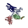 8iy9C 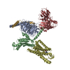 8iyhC 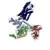 8jerC 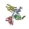 8jhnC M: map data used to model this data C: citing same article ( |
|---|---|
| Similar structure data |

-Links
 PDBj
PDBj


- Assembly
| Deposited unit | 
|
|---|---|
| 1 |
|
-Components
-Guanine nucleotide-binding protein ... , 3 types, 3 molecules BCG
| #1: Protein | Mass: 27024.762 Da / Num. of mol.: 1 / Mutation: G42D,E43N,A227D,G230D,I332A,V335I Source method: isolated from a genetically manipulated source Source: (gene. exp.) Homo sapiens (human) / Gene: GNAO1 / Production host:  Escherichia coli (E. coli) / References: UniProt: P09471 Escherichia coli (E. coli) / References: UniProt: P09471 |
|---|---|
| #2: Protein | Mass: 38534.062 Da / Num. of mol.: 1 Source method: isolated from a genetically manipulated source Source: (gene. exp.) Homo sapiens (human) / Gene: GNB1 / Production host:   Spodoptera frugiperda (fall armyworm) / References: UniProt: P62873 Spodoptera frugiperda (fall armyworm) / References: UniProt: P62873 |
| #3: Protein | Mass: 7861.143 Da / Num. of mol.: 1 Source method: isolated from a genetically manipulated source Source: (gene. exp.) Homo sapiens (human) / Gene: GNG2 / Production host: Spodoptera frugiperda (fall armyworm) / References: UniProt: P59768 |
-Antibody / Protein / Non-polymers , 3 types, 3 molecules HR
| #4: Antibody | Mass: 26466.486 Da / Num. of mol.: 1 Source method: isolated from a genetically manipulated source Source: (gene. exp.) Mus musculus (house mouse) / Production host: Escherichia coli (E. coli) |
|---|---|
| #5: Protein | Mass: 47798.926 Da / Num. of mol.: 1 Source method: isolated from a genetically manipulated source Source: (gene. exp.) Homo sapiens (human) / Gene: CHRM4, HCAR2 / Production host: Spodoptera frugiperda (fall armyworm) / References: UniProt: P08173, UniProt: Q8TDS4 |
| #6: Chemical | ChemComp-OKL /  Mass: 256.689 Da / Num. of mol.: 1 / Source method: obtained synthetically / Formula: C10H13ClN4O2 / Feature type: SUBJECT OF INVESTIGATION Mass: 256.689 Da / Num. of mol.: 1 / Source method: obtained synthetically / Formula: C10H13ClN4O2 / Feature type: SUBJECT OF INVESTIGATION |
-Details
| Has ligand of interest | Y |
|---|
-Experimental details
-Experiment
| Experiment | Method: ELECTRON MICROSCOPY |
|---|---|
| EM experiment | Aggregation state: PARTICLE / 3D reconstruction method: single particle reconstruction |
- Sample preparation
| Component |
| |||||||||||||||||||||||||||||||||||||||||||||||||
|---|---|---|---|---|---|---|---|---|---|---|---|---|---|---|---|---|---|---|---|---|---|---|---|---|---|---|---|---|---|---|---|---|---|---|---|---|---|---|---|---|---|---|---|---|---|---|---|---|---|---|
| Molecular weight | Experimental value: NO | |||||||||||||||||||||||||||||||||||||||||||||||||
| Source (natural) |
| |||||||||||||||||||||||||||||||||||||||||||||||||
| Source (recombinant) |
| |||||||||||||||||||||||||||||||||||||||||||||||||
| Buffer solution | pH: 7.4 | |||||||||||||||||||||||||||||||||||||||||||||||||
| Specimen | Embedding applied: NO / Shadowing applied: NO / Staining applied: NO / Vitrification applied: YES | |||||||||||||||||||||||||||||||||||||||||||||||||
| Vitrification | Cryogen name: ETHANE |
- Electron microscopy imaging
| Microscopy | Model: TFS GLACIOS |
|---|---|
| Electron gun | Electron source: FIELD EMISSION GUN / Accelerating voltage: 200 kV / Illumination mode: FLOOD BEAM |
| Electron lens | Mode: BRIGHT FIELDBright-field microscopy / Nominal defocus max: 2500 nm / Nominal defocus min: 500 nm |
| Specimen holder | Cryogen: NITROGEN |
| Image recording | Electron dose: 55 e/Å2 / Detector mode: COUNTING / Film or detector model: GATAN K3 BIOQUANTUM (6k x 4k) |
| Image scans | Movie frames/image: 40 |
- Processing
| EM software |
| ||||||||||||||||||||||||
|---|---|---|---|---|---|---|---|---|---|---|---|---|---|---|---|---|---|---|---|---|---|---|---|---|---|
| CTF correction | Type: NONE | ||||||||||||||||||||||||
| Particle selection | Num. of particles selected: 5761414 | ||||||||||||||||||||||||
| 3D reconstruction | Resolution: 3.45 Å / Resolution method: FSC 0.143 CUT-OFF / Num. of particles: 523816 / Symmetry type: POINT | ||||||||||||||||||||||||
| Atomic model building | Protocol: FLEXIBLE FIT / Space: REAL | ||||||||||||||||||||||||
| Atomic model building | PDB-ID: 8IY9 Accession code: 8IY9 / Source name: PDB / Type: experimental model | ||||||||||||||||||||||||
| Refine LS restraints |
|
