[English] 日本語
Yorodumi
- EMDB-36193: Structure of Acipimox-GPR109A-G protein complex -

+
Open data


ID or keywords:

Loading...

-
Basic information

Entry
Database: EMDB / ID: EMD-36193
TitleStructure of Acipimox-GPR109A-G protein complex
Map data
Sample
  • Complex: Acipimox-GPR109A-G protein complex
    • Complex: Guanine nucleotide-binding protein G(o) subunit alpha
      • Protein or peptide: Guanine nucleotide-binding protein G(o) subunit alpha
    • Complex: Guanine nucleotide-binding protein G(I)/G(S)/G(T) subunit beta-1
      • Protein or peptide: Guanine nucleotide-binding protein G(I)/G(S)/G(T) subunit beta-1
    • Complex: Guanine nucleotide-binding protein G(I)/G(S)/G(O) subunit gamma-2
      • Protein or peptide: Guanine nucleotide-binding protein G(I)/G(S)/G(O) subunit gamma-2
    • Complex: ScFv16 Antibody fragment
      • Protein or peptide: ScFv16 Antibody fragment
    • Complex: Hydroxycarboxylic acid receptor 2/GPR109A
      • Protein or peptide: Hydroxycarboxylic acid receptor 2
  • Ligand: 5-methyl-4-oxidanyl-pyrazin-4-ium-2-carboxylic acid
KeywordsGPCR / G protein / SIGNALING PROTEIN
Function / homology
Function and homology information


nicotinic acid receptor activity / Hydroxycarboxylic acid-binding receptors / Muscarinic acetylcholine receptors / neutrophil apoptotic process / G protein-coupled acetylcholine receptor activity / adenylate cyclase-inhibiting G protein-coupled acetylcholine receptor signaling pathway / Class A/1 (Rhodopsin-like receptors) / mu-type opioid receptor binding / corticotropin-releasing hormone receptor 1 binding / positive regulation of neutrophil apoptotic process ...nicotinic acid receptor activity / Hydroxycarboxylic acid-binding receptors / Muscarinic acetylcholine receptors / neutrophil apoptotic process / G protein-coupled acetylcholine receptor activity / adenylate cyclase-inhibiting G protein-coupled acetylcholine receptor signaling pathway / Class A/1 (Rhodopsin-like receptors) / mu-type opioid receptor binding / corticotropin-releasing hormone receptor 1 binding / positive regulation of neutrophil apoptotic process / G protein-coupled serotonin receptor activity / regulation of locomotion / positive regulation of adiponectin secretion / dopamine receptor signaling pathway / G protein-coupled receptor signaling pathway, coupled to cyclic nucleotide second messenger / G protein-coupled serotonin receptor binding / negative regulation of lipid catabolic process / muscle contraction / G-protein beta/gamma-subunit complex binding / Olfactory Signaling Pathway / Activation of the phototransduction cascade / G beta:gamma signalling through PLC beta / Presynaptic function of Kainate receptors / Thromboxane signalling through TP receptor / adenylate cyclase-modulating G protein-coupled receptor signaling pathway / G-protein activation / G protein-coupled acetylcholine receptor signaling pathway / Activation of G protein gated Potassium channels / Inhibition of voltage gated Ca2+ channels via Gbeta/gamma subunits / Prostacyclin signalling through prostacyclin receptor / Glucagon signaling in metabolic regulation / G beta:gamma signalling through CDC42 / ADP signalling through P2Y purinoceptor 12 / G beta:gamma signalling through BTK / Sensory perception of sweet, bitter, and umami (glutamate) taste / Synthesis, secretion, and inactivation of Glucagon-like Peptide-1 (GLP-1) / photoreceptor disc membrane / Adrenaline,noradrenaline inhibits insulin secretion / Glucagon-type ligand receptors / Vasopressin regulates renal water homeostasis via Aquaporins / G alpha (z) signalling events / cellular response to catecholamine stimulus / Glucagon-like Peptide-1 (GLP1) regulates insulin secretion / ADORA2B mediated anti-inflammatory cytokines production / adenylate cyclase-activating dopamine receptor signaling pathway / ADP signalling through P2Y purinoceptor 1 / G beta:gamma signalling through PI3Kgamma / cellular response to prostaglandin E stimulus / Cooperation of PDCL (PhLP1) and TRiC/CCT in G-protein beta folding / sensory perception of taste / GPER1 signaling / G-protein beta-subunit binding / heterotrimeric G-protein complex / Inactivation, recovery and regulation of the phototransduction cascade / extracellular vesicle / G alpha (12/13) signalling events / signaling receptor complex adaptor activity / Thrombin signalling through proteinase activated receptors (PARs) / retina development in camera-type eye / GTPase binding / cell junction / Ca2+ pathway / phospholipase C-activating G protein-coupled receptor signaling pathway / G alpha (i) signalling events / fibroblast proliferation / chemical synaptic transmission / G alpha (s) signalling events / postsynaptic membrane / G alpha (q) signalling events / Ras protein signal transduction / cell population proliferation / Extra-nuclear estrogen signaling / cell surface receptor signaling pathway / G protein-coupled receptor signaling pathway / lysosomal membrane / GTPase activity / dendrite / synapse / protein-containing complex binding / GTP binding / signal transduction / extracellular exosome / membrane / metal ion binding / plasma membrane / cytosol / cytoplasm
Similarity search - Function
Muscarinic acetylcholine receptor M4 / Muscarinic acetylcholine receptor family / G-protein alpha subunit, group I / Serpentine type 7TM GPCR chemoreceptor Srsx / G-alpha domain profile. / Guanine nucleotide binding protein (G-protein), alpha subunit / G protein alpha subunit, helical insertion / G-protein alpha subunit / G protein alpha subunit / G-protein, gamma subunit ...Muscarinic acetylcholine receptor M4 / Muscarinic acetylcholine receptor family / G-protein alpha subunit, group I / Serpentine type 7TM GPCR chemoreceptor Srsx / G-alpha domain profile. / Guanine nucleotide binding protein (G-protein), alpha subunit / G protein alpha subunit, helical insertion / G-protein alpha subunit / G protein alpha subunit / G-protein, gamma subunit / G-protein gamma subunit domain profile. / GGL domain / G-protein gamma-like domain superfamily / G-protein gamma-like domain / GGL domain / G protein gamma subunit-like motifs / Guanine nucleotide-binding protein, beta subunit / G-protein, beta subunit / G-protein coupled receptors family 1 signature. / G protein-coupled receptor, rhodopsin-like / GPCR, rhodopsin-like, 7TM / G-protein coupled receptors family 1 profile. / 7 transmembrane receptor (rhodopsin family) / G-protein beta WD-40 repeat / WD40 repeat, conserved site / Trp-Asp (WD) repeats signature. / WD domain, G-beta repeat / WD40 repeats / WD40 repeat / Trp-Asp (WD) repeats profile. / Trp-Asp (WD) repeats circular profile. / WD40-repeat-containing domain superfamily / WD40/YVTN repeat-like-containing domain superfamily / P-loop containing nucleoside triphosphate hydrolase
Similarity search - Domain/homology
Muscarinic acetylcholine receptor M4 / Guanine nucleotide-binding protein G(o) subunit alpha / Guanine nucleotide-binding protein G(I)/G(S)/G(O) subunit gamma-2 / Guanine nucleotide-binding protein G(I)/G(S)/G(T) subunit beta-1 / Hydroxycarboxylic acid receptor 2
Similarity search - Component
Biological speciesHomo sapiens (human) / Mus musculus (house mouse)
Methodsingle particle reconstruction / cryo EM / Resolution: 3.45 Å
AuthorsYadav MK / Sarma P / Chami M / Banerjee R / Shukla AK
Funding support India, 1 items
OrganizationGrant numberCountry
Department of Biotechnology (DBT, India)BT/PR29041/BRB/10/1697/20 India
Citation
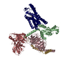
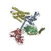
Journal: Nat Commun / Year: 2024
Title: Structure-guided engineering of biased-agonism in the human niacin receptor via single amino acid substitution.
Authors: Manish K Yadav / Parishmita Sarma / Jagannath Maharana / Manisankar Ganguly / Sudha Mishra / Nashrah Zaidi / Annu Dalal / Vinay Singh / Sayantan Saha / Gargi Mahajan / Saloni Sharma / ...Authors: Manish K Yadav / Parishmita Sarma / Jagannath Maharana / Manisankar Ganguly / Sudha Mishra / Nashrah Zaidi / Annu Dalal / Vinay Singh / Sayantan Saha / Gargi Mahajan / Saloni Sharma / Mohamed Chami / Ramanuj Banerjee / Arun K Shukla /
Abstract: The Hydroxycarboxylic acid receptor 2 (HCA2), also known as the niacin receptor or GPR109A, is a prototypical GPCR that plays a central role in the inhibition of lipolytic and atherogenic activities. ...The Hydroxycarboxylic acid receptor 2 (HCA2), also known as the niacin receptor or GPR109A, is a prototypical GPCR that plays a central role in the inhibition of lipolytic and atherogenic activities. Its activation also results in vasodilation that is linked to the side-effect of flushing associated with dyslipidemia drugs such as niacin. GPR109A continues to be a target for developing potential therapeutics in dyslipidemia with minimized flushing response. Here, we present cryo-EM structures of the GPR109A in complex with dyslipidemia drugs, niacin or acipimox, non-flushing agonists, MK6892 or GSK256073, and recently approved psoriasis drug, monomethyl fumarate (MMF). These structures elucidate the binding mechanism of agonists, molecular basis of receptor activation, and insights into biased signaling elicited by some of the agonists. The structural framework also allows us to engineer receptor mutants that exhibit G-protein signaling bias, and therefore, our study may help in structure-guided drug discovery efforts targeting this receptor.
History
DepositionMay 16, 2023-
Header (metadata) releaseMar 6, 2024-
Map releaseMar 6, 2024-
UpdateMar 20, 2024-
Current statusMar 20, 2024Processing site: PDBj / Status: Released

-
Structure visualization

Supplemental images

Downloads & links

-
Map

FileDownload / File: emd_36193.map.gz / Format: CCP4 / Size: 91.1 MB / Type: IMAGE STORED AS FLOATING POINT NUMBER (4 BYTES)
Voxel sizeX=Y=Z: 1.0975 Å
Density
Contour LevelBy AUTHOR: 0.14
Minimum - Maximum-1.4358217 - 2.5177112
Average (Standard dev.)0.0001201898 (±0.030356057)
SymmetrySpace group: 1
Details

EMDB XML:

Map geometry
Axis orderXYZ
Origin000
Dimensions288288288
Spacing288288288
CellA=B=C: 316.08 Å
α=β=γ: 90.0 °

-
Supplemental data

-
Half map: #2

Fileemd_36193_half_map_1.map
Projections & Slices
AxesZYX

Projections

Slices (1/2)
Density Histograms

-
Half map: #1

Fileemd_36193_half_map_2.map
Projections & Slices
AxesZYX

Projections

Slices (1/2)
Density Histograms

-
Sample components

+
Entire : Acipimox-GPR109A-G protein complex

EntireName: Acipimox-GPR109A-G protein complex
Components
  • Complex: Acipimox-GPR109A-G protein complex
    • Complex: Guanine nucleotide-binding protein G(o) subunit alpha
      • Protein or peptide: Guanine nucleotide-binding protein G(o) subunit alpha
    • Complex: Guanine nucleotide-binding protein G(I)/G(S)/G(T) subunit beta-1
      • Protein or peptide: Guanine nucleotide-binding protein G(I)/G(S)/G(T) subunit beta-1
    • Complex: Guanine nucleotide-binding protein G(I)/G(S)/G(O) subunit gamma-2
      • Protein or peptide: Guanine nucleotide-binding protein G(I)/G(S)/G(O) subunit gamma-2
    • Complex: ScFv16 Antibody fragment
      • Protein or peptide: ScFv16 Antibody fragment
    • Complex: Hydroxycarboxylic acid receptor 2/GPR109A
      • Protein or peptide: Hydroxycarboxylic acid receptor 2
  • Ligand: 5-methyl-4-oxidanyl-pyrazin-4-ium-2-carboxylic acid

+
Supramolecule #1: Acipimox-GPR109A-G protein complex

SupramoleculeName: Acipimox-GPR109A-G protein complex / type: complex / ID: 1 / Parent: 0 / Macromolecule list: #1-#2, #4-#5, #3
Source (natural)Organism: Homo sapiens (human)

+
Supramolecule #2: Guanine nucleotide-binding protein G(o) subunit alpha

SupramoleculeName: Guanine nucleotide-binding protein G(o) subunit alpha / type: complex / ID: 2 / Parent: 1 / Macromolecule list: #1
Details: This is a variant of Guanine nucleotide-binding protein G(o) subunit alpha called the "mini G(o) alpha"
Source (natural)Organism: Homo sapiens (human)

+
Supramolecule #3: Guanine nucleotide-binding protein G(I)/G(S)/G(T) subunit beta-1

SupramoleculeName: Guanine nucleotide-binding protein G(I)/G(S)/G(T) subunit beta-1
type: complex / ID: 3 / Parent: 1 / Macromolecule list: #2
Source (natural)Organism: Homo sapiens (human)

+
Supramolecule #4: Guanine nucleotide-binding protein G(I)/G(S)/G(O) subunit gamma-2

SupramoleculeName: Guanine nucleotide-binding protein G(I)/G(S)/G(O) subunit gamma-2
type: complex / ID: 4 / Parent: 1 / Macromolecule list: #4
Source (natural)Organism: Mus musculus (house mouse)

+
Supramolecule #5: ScFv16 Antibody fragment

SupramoleculeName: ScFv16 Antibody fragment / type: complex / ID: 5 / Parent: 1 / Macromolecule list: #5
Source (natural)Organism: Homo sapiens (human)

+
Supramolecule #6: Hydroxycarboxylic acid receptor 2/GPR109A

SupramoleculeName: Hydroxycarboxylic acid receptor 2/GPR109A / type: complex / ID: 6 / Parent: 1 / Macromolecule list: #3

+
Macromolecule #1: Guanine nucleotide-binding protein G(o) subunit alpha

MacromoleculeName: Guanine nucleotide-binding protein G(o) subunit alpha / type: protein_or_peptide / ID: 1 / Number of copies: 1 / Enantiomer: LEVO
Source (natural)Organism: Homo sapiens (human)
Molecular weightTheoretical: 27.024762 KDa
Recombinant expressionOrganism: Escherichia coli (E. coli)
SequenceString: MGHHHHHHEN LYFQGTLSAE ERAALERSKA IEKNLKEDGI SAAKDVKLLL LGADNSGKST IVKQMKIIHG GSGGSGGTTG IVETHFTFK NLHFRLFDVG GQRSERKKWI HCFEDVTAII FCVDLSDYNR MHESLMLFDS ICNNKFFIDT SIILFLNKKD L FGEKIKKS ...String:
MGHHHHHHEN LYFQGTLSAE ERAALERSKA IEKNLKEDGI SAAKDVKLLL LGADNSGKST IVKQMKIIHG GSGGSGGTTG IVETHFTFK NLHFRLFDVG GQRSERKKWI HCFEDVTAII FCVDLSDYNR MHESLMLFDS ICNNKFFIDT SIILFLNKKD L FGEKIKKS PLTICFPEYT GPNTYEDAAA YIQAQFESKN RSPNKEIYCH MTCATDTNNA QVIFDAVTDI IIANNLRGCG LY

UniProtKB: Guanine nucleotide-binding protein G(o) subunit alpha, Guanine nucleotide-binding protein G(o) subunit alpha

+
Macromolecule #2: Guanine nucleotide-binding protein G(I)/G(S)/G(T) subunit beta-1

MacromoleculeName: Guanine nucleotide-binding protein G(I)/G(S)/G(T) subunit beta-1
type: protein_or_peptide / ID: 2 / Number of copies: 1 / Enantiomer: LEVO
Source (natural)Organism: Homo sapiens (human)
Molecular weightTheoretical: 38.534062 KDa
Recombinant expressionOrganism: Spodoptera frugiperda (fall armyworm)
SequenceString: MHHHHHHGSS GSELDQLRQE AEQLKNQIRD ARKACADATL SQITNNIDPV GRIQMRTRRT LRGHLAKIYA MHWGTDSRLL VSASQDGKL IIWDSYTTNK VHAIPLRSSW VMTCAYAPSG NYVACGGLDN ICSIYNLKTR EGNVRVSREL AGHTGYLSCC R FLDDNQIV ...String:
MHHHHHHGSS GSELDQLRQE AEQLKNQIRD ARKACADATL SQITNNIDPV GRIQMRTRRT LRGHLAKIYA MHWGTDSRLL VSASQDGKL IIWDSYTTNK VHAIPLRSSW VMTCAYAPSG NYVACGGLDN ICSIYNLKTR EGNVRVSREL AGHTGYLSCC R FLDDNQIV TSSGDTTCAL WDIETGQQTT TFTGHTGDVM SLSLAPDTRL FVSGACDASA KLWDVREGMC RQTFTGHESD IN AICFFPN GNAFATGSDD ATCRLFDLRA DQELMTYSHD NIICGITSVS FSKSGRLLLA GYDDFNCNVW DALKADRAGV LAG HDNRVS CLGVTDDGMA VATGSWDSFL KIWN

UniProtKB: Guanine nucleotide-binding protein G(I)/G(S)/G(T) subunit beta-1

+
Macromolecule #3: Hydroxycarboxylic acid receptor 2

MacromoleculeName: Hydroxycarboxylic acid receptor 2 / type: protein_or_peptide / ID: 3 / Number of copies: 1 / Enantiomer: LEVO
Source (natural)Organism: Homo sapiens (human)
Molecular weightTheoretical: 47.798926 KDa
Recombinant expressionOrganism: Spodoptera frugiperda (fall armyworm)
SequenceString: MGKTIIALSY IFCLVFADYK DDDDAANFTP VNGSSGNQSV RLVTSSSLEV LFQGPGSNRH HLQDHFLEID KKNCCVFRDD FIVKVLPPV LGLEFIFGLL GNGLALWIFC FHLKSWKSSR IFLFNLAVAD FLLIICLPFL MDNYVRRWDW KFGDIPCRLM L FMLAMNRQ ...String:
MGKTIIALSY IFCLVFADYK DDDDAANFTP VNGSSGNQSV RLVTSSSLEV LFQGPGSNRH HLQDHFLEID KKNCCVFRDD FIVKVLPPV LGLEFIFGLL GNGLALWIFC FHLKSWKSSR IFLFNLAVAD FLLIICLPFL MDNYVRRWDW KFGDIPCRLM L FMLAMNRQ GSIIFLTVVA VDRYFRVVHP HHALNKISNR TAAIISCLLW GITIGLTVHL LKKKMPIQNG GANLCSSFSI CH TFQWHEA MFLLEFFLPL GIILFCSARI IWSLRQRQMD RHAKIKRAIT FIMVVAIVFV ICFLPSVVVR IRIFWLLHTS GTQ NCEVYR SVDLAFFITL SFTYMNSMLD PVVYYFSSPS FPNFFSTLIN RCLQRKMTGE PDNNRSTSVE LTGDPNKTRG APEA LMANS GEPWSPSYLG PTSP

UniProtKB: Muscarinic acetylcholine receptor M4, Hydroxycarboxylic acid receptor 2

+
Macromolecule #4: Guanine nucleotide-binding protein G(I)/G(S)/G(O) subunit gamma-2

MacromoleculeName: Guanine nucleotide-binding protein G(I)/G(S)/G(O) subunit gamma-2
type: protein_or_peptide / ID: 4 / Number of copies: 1 / Enantiomer: LEVO
Source (natural)Organism: Homo sapiens (human)
Molecular weightTheoretical: 7.861143 KDa
Recombinant expressionOrganism: Spodoptera frugiperda (fall armyworm)
SequenceString:
MASNNTASIA QARKLVEQLK MEANIDRIKV SKAAADLMAY CEAHAKEDPL LTPVPASENP FREKKFFCAI L

UniProtKB: Guanine nucleotide-binding protein G(I)/G(S)/G(O) subunit gamma-2

+
Macromolecule #5: ScFv16 Antibody fragment

MacromoleculeName: ScFv16 Antibody fragment / type: protein_or_peptide / ID: 5 / Number of copies: 1 / Enantiomer: LEVO
Source (natural)Organism: Mus musculus (house mouse)
Molecular weightTheoretical: 26.466486 KDa
Recombinant expressionOrganism: Escherichia coli (E. coli)
SequenceString: DVQLVESGGG LVQPGGSRKL SCSASGFAFS SFGMHWVRQA PEKGLEWVAY ISSGSGTIYY ADTVKGRFTI SRDDPKNTLF LQMTSLRSE DTAMYYCVRS IYYYGSSPFD FWGQGTTLTV SSGGGGSGGG GSGGGGSDIV MTQATSSVPV TPGESVSISC R SSKSLLHS ...String:
DVQLVESGGG LVQPGGSRKL SCSASGFAFS SFGMHWVRQA PEKGLEWVAY ISSGSGTIYY ADTVKGRFTI SRDDPKNTLF LQMTSLRSE DTAMYYCVRS IYYYGSSPFD FWGQGTTLTV SSGGGGSGGG GSGGGGSDIV MTQATSSVPV TPGESVSISC R SSKSLLHS NGNTYLYWFL QRPGQSPQLL IYRMSNLASG VPDRFSGSGS GTAFTLTISR LEAEDVGVYY CMQHLEYPLT FG AGTKLEL K

+
Macromolecule #6: 5-methyl-4-oxidanyl-pyrazin-4-ium-2-carboxylic acid

MacromoleculeName: 5-methyl-4-oxidanyl-pyrazin-4-ium-2-carboxylic acid / type: ligand / ID: 6 / Number of copies: 1 / Formula: OJX
Molecular weightTheoretical: 155.131 Da
Chemical component information

ChemComp-OJX:
5-methyl-4-oxidanyl-pyrazin-4-ium-2-carboxylic acid

-
Experimental details

-
Structure determination

Methodcryo EM
Processingsingle particle reconstruction
Aggregation stateparticle

-
Sample preparation

BufferpH: 7.4
VitrificationCryogen name: ETHANE

-
Electron microscopy

MicroscopeTFS GLACIOS
Electron beamAcceleration voltage: 200 kV / Electron source: FIELD EMISSION GUN
Electron opticsIllumination mode: FLOOD BEAM / Imaging mode: BRIGHT FIELDBright-field microscopy / Nominal defocus max: 2.5 µm / Nominal defocus min: 0.5 µm
Sample stageCooling holder cryogen: NITROGEN
Image recordingFilm or detector model: GATAN K3 BIOQUANTUM (6k x 4k) / Detector mode: COUNTING / Average electron dose: 52.0 e/Å2

-
Image processing

Particle selectionNumber selected: 9115816
Startup modelType of model: PDB ENTRY
PDB model - PDB ID:
Initial angle assignmentType: MAXIMUM LIKELIHOOD
Final 3D classificationSoftware - Name: cryoSPARC (ver. 3.3.2)
Final angle assignmentType: MAXIMUM LIKELIHOOD
Final reconstructionResolution.type: BY AUTHOR / Resolution: 3.45 Å / Resolution method: FSC 0.143 CUT-OFF / Software - Name: cryoSPARC (ver. 3.3.2) / Number images used: 1059994
FSC plot (resolution estimation)

-
Atomic model buiding 1

RefinementSpace: REAL / Protocol: FLEXIBLE FIT
Output model

PDB-8jer:
Structure of Acipimox-GPR109A-G protein complex

+
About Yorodumi

-
News

-
Feb 9, 2022. New format data for meta-information of EMDB entries

New format data for meta-information of EMDB entries

  • Version 3 of the EMDB header file is now the official format.
  • The previous official version 1.9 will be removed from the archive.

Related info.:EMDB header

External links:wwPDB to switch to version 3 of the EMDB data model

-
Aug 12, 2020. Covid-19 info

Covid-19 info

URL: https://pdbj.org/emnavi/covid19.php

New page: Covid-19 featured information page in EM Navigator.

Related info.:Covid-19 info / Mar 5, 2020. Novel coronavirus structure data

+
Mar 5, 2020. Novel coronavirus structure data

Novel coronavirus structure data

Related info.:Yorodumi Speices / Aug 12, 2020. Covid-19 info

External links:COVID-19 featured content - PDBj / Molecule of the Month (242):Coronavirus Proteases

+
Jan 31, 2019. EMDB accession codes are about to change! (news from PDBe EMDB page)

EMDB accession codes are about to change! (news from PDBe EMDB page)

  • The allocation of 4 digits for EMDB accession codes will soon come to an end. Whilst these codes will remain in use, new EMDB accession codes will include an additional digit and will expand incrementally as the available range of codes is exhausted. The current 4-digit format prefixed with “EMD-” (i.e. EMD-XXXX) will advance to a 5-digit format (i.e. EMD-XXXXX), and so on. It is currently estimated that the 4-digit codes will be depleted around Spring 2019, at which point the 5-digit format will come into force.
  • The EM Navigator/Yorodumi systems omit the EMD- prefix.

Related info.:Q: What is EMD? / ID/Accession-code notation in Yorodumi/EM Navigator

External links:EMDB Accession Codes are Changing Soon! / Contact to PDBj

+
Jul 12, 2017. Major update of PDB

Major update of PDB

  • wwPDB released updated PDB data conforming to the new PDBx/mmCIF dictionary.
  • This is a major update changing the version number from 4 to 5, and with Remediation, in which all the entries are updated.
  • In this update, many items about electron microscopy experimental information are reorganized (e.g. em_software).
  • Now, EM Navigator and Yorodumi are based on the updated data.

External links:wwPDB Remediation / Enriched Model Files Conforming to OneDep Data Standards Now Available in the PDB FTP Archive

-
Yorodumi

Thousand views of thousand structures

  • Yorodumi is a browser for structure data from EMDB, PDB, SASBDB, etc.
  • This page is also the successor to EM Navigator detail page, and also detail information page/front-end page for Omokage search.
  • The word "yorodu" (or yorozu) is an old Japanese word meaning "ten thousand". "mi" (miru) is to see.

Related info.:EMDB / PDB / SASBDB / Comparison of 3 databanks / Yorodumi Search / Aug 31, 2016. New EM Navigator & Yorodumi / Yorodumi Papers / Jmol/JSmol / Function and homology information / Changes in new EM Navigator and Yorodumi

Read more