 Movie
Movie Controller

[English] 日本語
 Yorodumi
- PDB-8hx6: Crystal structure of 4-amino-4-deoxychorismate synthase from Stre... -
+ Open data

- Basic information
| Entry | Database: PDB / ID: 8hx6 | ||||||
|---|---|---|---|---|---|---|---|
| Title | Crystal structure of 4-amino-4-deoxychorismate synthase from Streptomyces venezuelae | ||||||
 Components Components | 4-amino-4-deoxychorismate synthase | ||||||
 Keywords Keywords |  TRANSFERASE / Synthase / Multi-domain / Chorismate TRANSFERASE / Synthase / Multi-domain / Chorismate | ||||||
| Function / homology |  Function and homology informationaminodeoxychorismate synthase / 4-amino-4-deoxychorismate synthase activity / folic acid-containing compound biosynthetic process / glutamine metabolic process Function and homology informationaminodeoxychorismate synthase / 4-amino-4-deoxychorismate synthase activity / folic acid-containing compound biosynthetic process / glutamine metabolic processSimilarity search - Function | ||||||
| Biological species |  Streptomyces venezuelae (bacteria) Streptomyces venezuelae (bacteria) | ||||||
| Method | X-RAY DIFFRACTION / SYNCHROTRON / MOLECULAR REPLACEMENT / Resolution: 2.14 Å | ||||||
 Authors Authors | Nakamichi, Y. / Watanabe, M. | ||||||
| Funding support |  Japan, 1items Japan, 1items
| ||||||
 Citation Citation | Journal: Acta Crystallogr D Struct Biol / Year: 2023 Title: Structural basis for the allosteric pathway of 4-amino-4-deoxychorismate synthase. Authors: Nakamichi, Y. / Kobayashi, J. / Toyoda, K. / Suda, M. / Hiraga, K. / Inui, M. / Watanabe, M. | ||||||
| History |
|
- Structure visualization
| Structure viewer | Molecule: MolmilJmol/JSmol |
|---|
- Downloads & links
-Download
| PDBx/mmCIF format | 8hx6.cif.gz | 518.7 KB | Display | PDBx/mmCIF format |
|---|---|---|---|---|
| PDB format | pdb8hx6.ent.gz | 427.1 KB | Display | PDB format |
| PDBx/mmJSON format | 8hx6.json.gz | Tree view | PDBx/mmJSON format | |
| Others |  Other downloads Other downloads |
-Validation report
| Arichive directory | https://data.pdbj.org/pub/pdb/validation_reports/hx/8hx6ftp://data.pdbj.org/pub/pdb/validation_reports/hx/8hx6 | HTTPS FTP |
|---|
-Related structure data
| Related structure data | 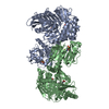 8hx7C 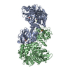 8hx8C 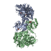 8hx9C 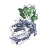 1qdlS S: Starting model for refinement C: citing same article ( |
|---|---|
| Similar structure data |
-Links
 PDBj
PDBj


- Assembly
| Deposited unit | 
| ||||||||
|---|---|---|---|---|---|---|---|---|---|
| 1 |
| ||||||||
| Unit cell |
|
-Components
-Protein , 1 types, 2 molecules AB
| #1: Protein | Mass: 76744.352 Da / Num. of mol.: 2 Source method: isolated from a genetically manipulated source Source: (gene. exp.) Streptomyces venezuelae (bacteria) / Gene: papA / Plasmid: pColdI / Production host: Escherichia coli BL21(DE3) (bacteria) / References: UniProt: Q6L8Q5, aminodeoxychorismate synthase |
|---|
-Non-polymers , 5 types, 56 molecules 


| #2: Chemical | Tryptophan Type: L-peptide linking / Mass: 204.225 Da / Num. of mol.: 2 / Source method: obtained synthetically / Formula: C11H12N2O2 / Feature type: SUBJECT OF INVESTIGATION Type: L-peptide linking / Mass: 204.225 Da / Num. of mol.: 2 / Source method: obtained synthetically / Formula: C11H12N2O2 / Feature type: SUBJECT OF INVESTIGATION#3: Chemical | ChemComp-MLT / | Malic acid Mass: 134.087 Da / Num. of mol.: 1 / Source method: obtained synthetically / Formula: C4H6O5 Mass: 134.087 Da / Num. of mol.: 1 / Source method: obtained synthetically / Formula: C4H6O5#4: Chemical | ChemComp-MG / |  Mass: 24.305 Da / Num. of mol.: 1 / Source method: obtained synthetically / Formula: Mg / Feature type: SUBJECT OF INVESTIGATION Mass: 24.305 Da / Num. of mol.: 1 / Source method: obtained synthetically / Formula: Mg / Feature type: SUBJECT OF INVESTIGATION#5: Chemical | ChemComp-SIN / | Succinic acid Mass: 118.088 Da / Num. of mol.: 1 / Source method: obtained synthetically / Formula: C4H6O4 Mass: 118.088 Da / Num. of mol.: 1 / Source method: obtained synthetically / Formula: C4H6O4#6: Water | ChemComp-HOH / | WaterMass: 18.015 Da / Num. of mol.: 51 / Source method: isolated from a natural source / Formula: H2O |
|---|
-Details
| Has ligand of interest | Y |
|---|
-Experimental details
-Experiment
| Experiment | Method: X-RAY DIFFRACTION / Number of used crystals: 1 |
|---|
- Sample preparation
| Crystal | Density Matthews: 2.22 Å3/Da / Density % sol: 44.48 % |
|---|---|
| Crystal grow | Temperature: 293 K / Method: vapor diffusion, hanging drop / pH: 7 Details: 51% (v/v) Tacsimate, 0.1 M Bis-tris propane (pH 7.0) |
-Data collection
| Diffraction | Mean temperature: 100 K / Serial crystal experiment: N |
|---|---|
| Diffraction source | Source: SYNCHROTRON / Site: SPring-8 / Beamline: BL44XU / Wavelength: 0.9 Å |
| Detector | Type: DECTRIS EIGER X 16M / Detector: PIXEL / Date: Jul 3, 2020 |
| Radiation | Protocol: SINGLE WAVELENGTH / Monochromatic (M) / Laue (L): M / Scattering type: x-ray |
| Radiation wavelength | Wavelength: 0.9 Å / Relative weight: 1 |
| Reflection | Resolution: 2.14→47.27 Å / Num. obs: 74670 / % possible obs: 99.8 % / Redundancy: 6.8 % / Biso Wilson estimate: 52.19 Å2 / CC1/2: 0.999 / Rpim(I) all: 0.023 / Net I/σ(I): 18.99 |
| Reflection shell | Resolution: 2.14→2.22 Å / Redundancy: 6.7 % / Mean I/σ(I) obs: 1.29 / Num. unique obs: 7224 / CC1/2: 0.64 / Rpim(I) all: 0.581 / % possible all: 98.1 |
- Processing
| Software |
| ||||||||||||||||||||||||||||||||||||||||||||||||||||||||||||||||||||||||||||||||||||||||||||||||||||||||||||||||||||||||||||||||||||||||||||||||||||||||||||||||||||||||||||||||||||||
|---|---|---|---|---|---|---|---|---|---|---|---|---|---|---|---|---|---|---|---|---|---|---|---|---|---|---|---|---|---|---|---|---|---|---|---|---|---|---|---|---|---|---|---|---|---|---|---|---|---|---|---|---|---|---|---|---|---|---|---|---|---|---|---|---|---|---|---|---|---|---|---|---|---|---|---|---|---|---|---|---|---|---|---|---|---|---|---|---|---|---|---|---|---|---|---|---|---|---|---|---|---|---|---|---|---|---|---|---|---|---|---|---|---|---|---|---|---|---|---|---|---|---|---|---|---|---|---|---|---|---|---|---|---|---|---|---|---|---|---|---|---|---|---|---|---|---|---|---|---|---|---|---|---|---|---|---|---|---|---|---|---|---|---|---|---|---|---|---|---|---|---|---|---|---|---|---|---|---|---|---|---|---|---|
| Refinement | Method to determine structure: MOLECULAR REPLACEMENT Starting model: 1qdl Resolution: 2.14→47.26 Å / Cor.coef. Fo:Fc: 0.958 / Cor.coef. Fo:Fc free: 0.932 / SU B: 25.22 / SU ML: 0.268 / Cross valid method: THROUGHOUT / ESU R: 0.286 / ESU R Free: 0.225 / Stereochemistry target values: MAXIMUM LIKELIHOOD / Details: HYDROGENS HAVE BEEN ADDED IN THE RIDING POSITIONS
| ||||||||||||||||||||||||||||||||||||||||||||||||||||||||||||||||||||||||||||||||||||||||||||||||||||||||||||||||||||||||||||||||||||||||||||||||||||||||||||||||||||||||||||||||||||||
| Solvent computation | Ion probe radii: 0.8 Å / Shrinkage radii: 0.8 Å / VDW probe radii: 1.2 Å / Solvent model: MASK | ||||||||||||||||||||||||||||||||||||||||||||||||||||||||||||||||||||||||||||||||||||||||||||||||||||||||||||||||||||||||||||||||||||||||||||||||||||||||||||||||||||||||||||||||||||||
| Displacement parameters | Biso mean: 64.996 Å2
| ||||||||||||||||||||||||||||||||||||||||||||||||||||||||||||||||||||||||||||||||||||||||||||||||||||||||||||||||||||||||||||||||||||||||||||||||||||||||||||||||||||||||||||||||||||||
| Refinement step | Cycle: 1 / Resolution: 2.14→47.26 Å
| ||||||||||||||||||||||||||||||||||||||||||||||||||||||||||||||||||||||||||||||||||||||||||||||||||||||||||||||||||||||||||||||||||||||||||||||||||||||||||||||||||||||||||||||||||||||
| Refine LS restraints |
|