 Movie
Movie Controller


+ Open data

- Basic information
| Entry | Database: PDB / ID: 8htg | ||||||
|---|---|---|---|---|---|---|---|
| Title | Crystal structure of Golf in complex with GTP-gamma S and Mg | ||||||
 Components Components | Guanine nucleotide-binding protein G(olf) subunit alpha | ||||||
 Keywords Keywords |  SIGNALING PROTEIN / G protein SIGNALING PROTEIN / G protein | ||||||
| Function / homology |  Function and homology information Function and homology informationAdenylate cyclase activating pathway / Adenylate cyclase inhibitory pathway / Olfactory Signaling Pathway / cellular response to dopamine / response to caffeine / regulation of long-term synaptic depression / response to amphetamine / G protein-coupled receptor binding / G-protein beta/gamma-subunit complex binding / adenylate cyclase-modulating G protein-coupled receptor signaling pathway ...Adenylate cyclase activating pathway / Adenylate cyclase inhibitory pathway / Olfactory Signaling Pathway / cellular response to dopamine / response to caffeine / regulation of long-term synaptic depression / response to amphetamine / G protein-coupled receptor binding / G-protein beta/gamma-subunit complex binding / adenylate cyclase-modulating G protein-coupled receptor signaling pathway / adenylate cyclase-activating G protein-coupled receptor signaling pathway / adenylate cyclase-activating dopamine receptor signaling pathway / heterotrimeric G-protein complex / sensory perception of smell / G protein-coupled receptor signaling pathway / GTPase activity / GTP binding / metal ion binding / plasma membraneSimilarity search - Function | ||||||
| Biological species |  Mus musculus (house mouse) Mus musculus (house mouse) | ||||||
| Method | X-RAY DIFFRACTION / SYNCHROTRON / MOLECULAR REPLACEMENT / Resolution: 2.91 Å | ||||||
 Authors Authors | Kang, H. / Choi, H.-J. | ||||||
| Funding support |  Korea, Republic Of, 1items Korea, Republic Of, 1items
| ||||||
 Citation Citation | Journal: Nat Commun / Year: 2023 Title: Understanding the molecular mechanisms of odorant binding and activation of the human OR52 family. Authors: Chulwon Choi / Jungnam Bae / Seonghan Kim / Seho Lee / Hyunook Kang / Jinuk Kim / Injin Bang / Kiheon Kim / Won-Ki Huh / Chaok Seok / Hahnbeom Park / Wonpil Im / Hee-Jung Choi /  Abstract: Structural and mechanistic studies on human odorant receptors (ORs), key in olfactory signaling, are challenging because of their low surface expression in heterologous cells. The recent structure of ...Structural and mechanistic studies on human odorant receptors (ORs), key in olfactory signaling, are challenging because of their low surface expression in heterologous cells. The recent structure of OR51E2 bound to propionate provided molecular insight into odorant recognition, but the lack of an inactive OR structure limited understanding of the activation mechanism of ORs upon odorant binding. Here, we determined the cryo-electron microscopy structures of consensus OR52 (OR52), a representative of the OR52 family, in the ligand-free (apo) and octanoate-bound states. The apo structure of OR52 reveals a large opening between transmembrane helices (TMs) 5 and 6. A comparison between the apo and active structures of OR52 demonstrates the inward and outward movements of the extracellular and intracellular segments of TM6, respectively. These results, combined with molecular dynamics simulations and signaling assays, shed light on the molecular mechanisms of odorant binding and activation of the OR52 family. | ||||||
| History |
|
- Structure visualization
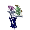
| Structure viewer | Molecule: MolmilJmol/JSmol |
|---|
- Downloads & links
-Download
| PDBx/mmCIF format | 8htg.cif.gz | 285 KB | Display | PDBx/mmCIF format |
|---|---|---|---|---|
| PDB format | pdb8htg.ent.gz | 227.3 KB | Display | PDB format |
| PDBx/mmJSON format | 8htg.json.gz | Tree view | PDBx/mmJSON format | |
| Others |  Other downloads Other downloads |
-Validation report
| Arichive directory | https://data.pdbj.org/pub/pdb/validation_reports/ht/8htgftp://data.pdbj.org/pub/pdb/validation_reports/ht/8htg | HTTPS FTP |
|---|
-Related structure data


-Links
 PDBj
PDBj


- Assembly
| Deposited unit | 
| ||||||||
|---|---|---|---|---|---|---|---|---|---|
| 1 |
| ||||||||
| 2 |
| ||||||||
| 3 |
| ||||||||
| 4 |
| ||||||||
| Unit cell |
|
-Components
-Protein , 1 types, 4 molecules ABCD
| #1: Protein | Mass: 46067.273 Da / Num. of mol.: 4 / Mutation: C3S Source method: isolated from a genetically manipulated source Source: (gene. exp.) Mus musculus (house mouse) / Gene: Gnal / Production host:   Spodoptera frugiperda (fall armyworm) / References: UniProt: Q8CGK7 Spodoptera frugiperda (fall armyworm) / References: UniProt: Q8CGK7 |
|---|
-Non-polymers , 5 types, 65 molecules 


| #2: Chemical | Ethylene glycol Mass: 62.068 Da / Num. of mol.: 2 / Source method: obtained synthetically / Formula: C2H6O2 Mass: 62.068 Da / Num. of mol.: 2 / Source method: obtained synthetically / Formula: C2H6O2#3: Chemical | ChemComp-MG /  Mass: 24.305 Da / Num. of mol.: 4 / Source method: obtained synthetically / Formula: Mg / Feature type: SUBJECT OF INVESTIGATION Mass: 24.305 Da / Num. of mol.: 4 / Source method: obtained synthetically / Formula: Mg / Feature type: SUBJECT OF INVESTIGATION#4: Chemical | ChemComp-GSP /  Mass: 539.246 Da / Num. of mol.: 4 / Source method: obtained synthetically / Formula: C10H16N5O13P3S / Feature type: SUBJECT OF INVESTIGATION Mass: 539.246 Da / Num. of mol.: 4 / Source method: obtained synthetically / Formula: C10H16N5O13P3S / Feature type: SUBJECT OF INVESTIGATION#5: Chemical | Phosphate Mass: 94.971 Da / Num. of mol.: 3 / Source method: obtained synthetically / Formula: PO4 Mass: 94.971 Da / Num. of mol.: 3 / Source method: obtained synthetically / Formula: PO4#6: Water | ChemComp-HOH / | WaterMass: 18.015 Da / Num. of mol.: 52 / Source method: isolated from a natural source / Formula: H2O |
|---|
-Details
| Has ligand of interest | Y |
|---|
-Experimental details
-Experiment
| Experiment | Method: X-RAY DIFFRACTION / Number of used crystals: 1 |
|---|
- Sample preparation
| Crystal | Density Matthews: 1.91 Å3/Da / Density % sol: 35.5 % |
|---|---|
| Crystal grow | Temperature: 295 K / Method: vapor diffusion, hanging drop / Details: 18% PEG 3350, 0.2M MgCl2 |
-Data collection
| Diffraction | Mean temperature: 100 K / Serial crystal experiment: N |
|---|---|
| Diffraction source | Source: SYNCHROTRON / Site: PAL/PLS / Beamline: 5C (4A) / Wavelength: 0.9794 Å |
| Detector | Type: DECTRIS EIGER X 9M / Detector: PIXEL / Date: May 21, 2020 |
| Radiation | Protocol: SINGLE WAVELENGTH / Monochromatic (M) / Laue (L): M / Scattering type: x-ray |
| Radiation wavelength | Wavelength: 0.9794 Å / Relative weight: 1 |
| Reflection | Resolution: 2.91→27.57 Å / Num. obs: 30932 / % possible obs: 99.4 % / Redundancy: 3.4 % / Biso Wilson estimate: 43.54 Å2 / CC1/2: 0.983 / CC star: 0.996 / Rmerge(I) obs: 0.1779 / Rpim(I) all: 0.1122 / Rrim(I) all: 0.2108 / Net I/σ(I): 7.13 |
| Reflection shell | Resolution: 2.91→3.01 Å / Redundancy: 3.5 % / Rmerge(I) obs: 0.8078 / Mean I/σ(I) obs: 1.48 / Num. unique obs: 3030 / CC1/2: 0.67 / CC star: 0.896 / Rpim(I) all: 0.5036 / Rrim(I) all: 0.9542 / % possible all: 99.84 |
- Processing
| Software |
| ||||||||||||||||||||||||||||||||||||||||||||||||||||||||||||||||||||||||||||||||||||
|---|---|---|---|---|---|---|---|---|---|---|---|---|---|---|---|---|---|---|---|---|---|---|---|---|---|---|---|---|---|---|---|---|---|---|---|---|---|---|---|---|---|---|---|---|---|---|---|---|---|---|---|---|---|---|---|---|---|---|---|---|---|---|---|---|---|---|---|---|---|---|---|---|---|---|---|---|---|---|---|---|---|---|---|---|---|
| Refinement | Method to determine structure: MOLECULAR REPLACEMENT / Resolution: 2.91→27.57 Å / SU ML: 0.42 / Cross valid method: FREE R-VALUE / σ(F): 1.36 / Phase error: 28.73 / Stereochemistry target values: ML
| ||||||||||||||||||||||||||||||||||||||||||||||||||||||||||||||||||||||||||||||||||||
| Solvent computation | Shrinkage radii: 0.9 Å / VDW probe radii: 1.11 Å / Solvent model: FLAT BULK SOLVENT MODEL | ||||||||||||||||||||||||||||||||||||||||||||||||||||||||||||||||||||||||||||||||||||
| Refinement step | Cycle: LAST / Resolution: 2.91→27.57 Å
| ||||||||||||||||||||||||||||||||||||||||||||||||||||||||||||||||||||||||||||||||||||
| Refine LS restraints |
| ||||||||||||||||||||||||||||||||||||||||||||||||||||||||||||||||||||||||||||||||||||
| LS refinement shell |
|
