 Movie
Movie Controller


+ Open data

- Basic information
| Entry | Database: PDB / ID: 8hqe | |||||||||
|---|---|---|---|---|---|---|---|---|---|---|
| Title | Cryo-EM structure of the apo-GPR132-Gi | |||||||||
 Components Components |
| |||||||||
 Keywords Keywords |  MEMBRANE PROTEIN / GPCR / GPR132 MEMBRANE PROTEIN / GPCR / GPR132 | |||||||||
| Function / homology |  Function and homology information Function and homology informationnegative regulation of G2/M transition of mitotic cell cycle / Class A/1 (Rhodopsin-like receptors) / Adenylate cyclase inhibitory pathway / positive regulation of protein localization to cell cortex / regulation of cAMP-mediated signaling / D2 dopamine receptor binding / G protein-coupled serotonin receptor binding / regulation of mitotic spindle organization / cellular response to forskolin / adenylate cyclase-inhibiting G protein-coupled receptor signaling pathway ...negative regulation of G2/M transition of mitotic cell cycle / Class A/1 (Rhodopsin-like receptors) / Adenylate cyclase inhibitory pathway / positive regulation of protein localization to cell cortex / regulation of cAMP-mediated signaling / D2 dopamine receptor binding / G protein-coupled serotonin receptor binding / regulation of mitotic spindle organization / cellular response to forskolin / adenylate cyclase-inhibiting G protein-coupled receptor signaling pathway / Regulation of insulin secretion / G protein-coupled receptor binding / G protein-coupled receptor activity / G1/S transition of mitotic cell cycle / Olfactory Signaling Pathway / G-protein beta/gamma-subunit complex binding / Activation of the phototransduction cascade / G beta:gamma signalling through PLC beta / Presynaptic function of Kainate receptors / Thromboxane signalling through TP receptor / adenylate cyclase-modulating G protein-coupled receptor signaling pathway / G-protein activation / G protein-coupled acetylcholine receptor signaling pathway / Activation of G protein gated Potassium channels / Inhibition of voltage gated Ca2+ channels via Gbeta/gamma subunits / Prostacyclin signalling through prostacyclin receptor / Glucagon signaling in metabolic regulation / G beta:gamma signalling through CDC42 / ADP signalling through P2Y purinoceptor 12 / G beta:gamma signalling through BTK / Synthesis, secretion, and inactivation of Glucagon-like Peptide-1 (GLP-1) / Sensory perception of sweet, bitter, and umami (glutamate) taste / photoreceptor disc membrane / response to peptide hormone / Adrenaline,noradrenaline inhibits insulin secretion / Glucagon-type ligand receptors / Vasopressin regulates renal water homeostasis via Aquaporins / G alpha (z) signalling events / cellular response to catecholamine stimulus / Glucagon-like Peptide-1 (GLP1) regulates insulin secretion / ADORA2B mediated anti-inflammatory cytokines production / adenylate cyclase-activating dopamine receptor signaling pathway / ADP signalling through P2Y purinoceptor 1 / G beta:gamma signalling through PI3Kgamma / cellular response to prostaglandin E stimulus / Cooperation of PDCL (PhLP1) and TRiC/CCT in G-protein beta folding / sensory perception of taste / GPER1 signaling / G-protein beta-subunit binding / GDP binding / Inactivation, recovery and regulation of the phototransduction cascade / heterotrimeric G-protein complex / G alpha (12/13) signalling events / extracellular vesicle / signaling receptor complex adaptor activity / Thrombin signalling through proteinase activated receptors (PARs) / retina development in camera-type eye / GTPase binding / Ca2+ pathway / phospholipase C-activating G protein-coupled receptor signaling pathway / cell cortex / midbody / G alpha (i) signalling events / fibroblast proliferation / G alpha (s) signalling events / G alpha (q) signalling events / Ras protein signal transduction / cell population proliferation / Extra-nuclear estrogen signaling / electron transfer activity / periplasmic space / cell cycle / iron ion binding / G protein-coupled receptor signaling pathway / lysosomal membrane / cell division / GTPase activity / centrosome / synapse / heme binding / protein-containing complex binding / GTP binding / nucleolus / magnesium ion binding / signal transduction / extracellular exosome / nucleoplasm / membrane / plasma membrane / cytosol / cytoplasmSimilarity search - Function | |||||||||
| Biological species |  Homo sapiens (human) Homo sapiens (human) Mus musculus (house mouse) Mus musculus (house mouse) Escherichia coli (E. coli) Escherichia coli (E. coli) | |||||||||
| Method | ELECTRON MICROSCOPY / single particle reconstruction / cryo EM / Resolution: 2.97 Å | |||||||||
 Authors Authors | Wang, J.L. / Ding, J.H. / Sun, J.P. / Yu, X. | |||||||||
| Funding support |  China, 2items China, 2items
| |||||||||
 Citation Citation | Journal: Nat Metab / Year: 2023 Title: Functional screening and rational design of compounds targeting GPR132 to treat diabetes. Authors: Jia-Le Wang / Xiao-Dong Dou / Jie Cheng / Ming-Xin Gao / Guo-Feng Xu / Wei Ding / Jin-Hui Ding / Yu Li / Si-Han Wang / Zhao-Wei Ji / Xin-Yi Zhao / Tong-Yu Huo / Cai-Fang Zhang / Ya-Meng Liu ...Authors: Jia-Le Wang / Xiao-Dong Dou / Jie Cheng / Ming-Xin Gao / Guo-Feng Xu / Wei Ding / Jin-Hui Ding / Yu Li / Si-Han Wang / Zhao-Wei Ji / Xin-Yi Zhao / Tong-Yu Huo / Cai-Fang Zhang / Ya-Meng Liu / Xue-Ying Sha / Jia-Rui Gao / Wen-Hui Zhang / Yong Hao / Cheng Zhang / Jin-Peng Sun / Ning Jiao / Xiao Yu / Abstract: Chronic inflammation due to islet-residing macrophages plays key roles in the development of type 2 diabetes mellitus. By systematically profiling intra-islet lipid-transmembrane receptor signalling ...Chronic inflammation due to islet-residing macrophages plays key roles in the development of type 2 diabetes mellitus. By systematically profiling intra-islet lipid-transmembrane receptor signalling in islet-resident macrophages, we identified endogenous 9(S)-hydroxy-10,12-octadecadienoic acid-G-protein-coupled receptor 132 (GPR132)-Gi signalling as a significant contributor to islet macrophage reprogramming and found that GPR132 deficiency in macrophages reversed metabolic disorders in mice fed a high-fat diet. The cryo-electron microscopy structures of GPR132 bound with two endogenous agonists, N-palmitoylglycine and 9(S)-hydroxy-10,12-octadecadienoic acid, enabled us to rationally design both GPR132 agonists and antagonists with high potency and selectivity through stepwise translational approaches. We ultimately identified a selective GPR132 antagonist, NOX-6-18, that modulates macrophage reprogramming within pancreatic islets, decreases weight gain and enhances glucose metabolism in mice fed a high-fat diet. Our study not only illustrates that intra-islet lipid signalling contributes to islet macrophage reprogramming but also provides a broadly applicable strategy for the identification of important G-protein-coupled receptor targets in pathophysiological processes, followed by the rational design of therapeutic leads for refractory diseases such as diabetes. | |||||||||
| History |
|
- Structure visualization
| Structure viewer | Molecule: MolmilJmol/JSmol |
|---|
- Downloads & links
-Download
| PDBx/mmCIF format | 8hqe.cif.gz | 237.1 KB | Display | PDBx/mmCIF format |
|---|---|---|---|---|
| PDB format | pdb8hqe.ent.gz | 180.4 KB | Display | PDB format |
| PDBx/mmJSON format | 8hqe.json.gz | Tree view | PDBx/mmJSON format | |
| Others |  Other downloads Other downloads |
-Validation report
| Arichive directory | https://data.pdbj.org/pub/pdb/validation_reports/hq/8hqeftp://data.pdbj.org/pub/pdb/validation_reports/hq/8hqe | HTTPS FTP |
|---|
-Related structure data
| Related structure data |  34948MC  8hqmC  8hqnC  8hviC M: map data used to model this data C: citing same article ( |
|---|---|
| Similar structure data |


-Links
 PDBj
PDBj


- Assembly
| Deposited unit | 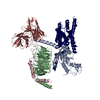
|
|---|---|
| 1 |
|
-Components
| #1: Protein | Mass: 39489.160 Da / Num. of mol.: 1 Source method: isolated from a genetically manipulated source Source: (gene. exp.) Homo sapiens (human) / Gene: GNB1 / Production host:  Baculovirus expression vector pFastBac1-HM / References: UniProt: P62873 Baculovirus expression vector pFastBac1-HM / References: UniProt: P62873 |
|---|---|
| #2: Protein | Mass: 6375.332 Da / Num. of mol.: 1 Source method: isolated from a genetically manipulated source Source: (gene. exp.) Homo sapiens (human) / Gene: GNG2 / Production host: Baculovirus expression vector pFastBac1-HM / References: UniProt: P59768 |
| #3: Antibody | Mass: 26610.615 Da / Num. of mol.: 1 Source method: isolated from a genetically manipulated source Source: (gene. exp.) Mus musculus (house mouse) / Production host: Baculovirus expression vector pFastBac1-HM |
| #4: Protein | Mass: 57125.316 Da / Num. of mol.: 1 Source method: isolated from a genetically manipulated source Source: (gene. exp.) Escherichia coli (E. coli), (gene. exp.) Homo sapiens (human)Gene: cybC, GPR132, G2A / Production host: Baculovirus expression vector pFastBac1-HM / References: UniProt: P0ABE7, UniProt: Q9UNW8 |
| #5: Protein | Mass: 40445.059 Da / Num. of mol.: 1 Source method: isolated from a genetically manipulated source Source: (gene. exp.) Homo sapiens (human) / Gene: GNAI1 / Production host: Baculovirus expression vector pFastBac1-HM / References: UniProt: P63096 |
-Experimental details
-Experiment
| Experiment | Method: ELECTRON MICROSCOPY |
|---|---|
| EM experiment | Aggregation state: PARTICLE / 3D reconstruction method: single particle reconstruction |
- Sample preparation
| Component | Name: Cryo-EM structure of the apo-GPR132-Gi / Type: COMPLEX / Entity ID: all / Source: MULTIPLE SOURCES |
|---|---|
| Molecular weight | Value: 160 kDa/nm / Experimental value: YES |
| Source (natural) | Organism: Homo sapiens (human) |
| Source (recombinant) | Organism: Baculovirus expression vector pFastBac1-HM |
| Buffer solution | pH: 7.5 |
| Specimen | Conc.: 5 mg/ml / Embedding applied: NO / Shadowing applied: NO / Staining applied: NO / Vitrification applied: YES |
| Vitrification | Cryogen name: ETHANE |
- Electron microscopy imaging
| Experimental equipment |  Model: Titan Krios / Image courtesy: FEI Company |
|---|---|
| Microscopy | Model: FEI TITAN KRIOS |
| Electron gun | Electron source: FIELD EMISSION GUN / Accelerating voltage: 300 kV / Illumination mode: SPOT SCAN |
| Electron lens | Mode: BRIGHT FIELDBright-field microscopy / Nominal defocus max: 2500 nm / Nominal defocus min: 1000 nm |
| Image recording | Electron dose: 60 e/Å2 / Film or detector model: GATAN K2 SUMMIT (4k x 4k) |
- Processing
| Software | Name: PHENIX / Version: 1.20.1_4487: / Classification: refinement |
|---|---|
| CTF correction | Type: PHASE FLIPPING AND AMPLITUDE CORRECTION |
| 3D reconstruction | Resolution: 2.97 Å / Resolution method: FSC 0.143 CUT-OFF / Num. of particles: 363528 / Symmetry type: POINT |
