 Movie
Movie Controller

[English] 日本語
 Yorodumi
- PDB-8hfo: Crystal Structure of Mycobacterium smegmatis MshC in Complex with... -
+ Open data

- Basic information
| Entry | Database: PDB / ID: 8hfo | |||||||||
|---|---|---|---|---|---|---|---|---|---|---|
| Title | Crystal Structure of Mycobacterium smegmatis MshC in Complex with Compound 7d | |||||||||
 Components Components | L-cysteine:1D-myo-inositol 2-amino-2-deoxy-alpha-D-glucopyranoside ligase | |||||||||
 Keywords Keywords | LIGASE / Rossmann fold / Inhibitor | |||||||||
| Function / homology |  Function and homology informationL-cysteine:1D-myo-inositol 2-amino-2-deoxy-alpha-D-glucopyranoside ligase / cysteine-glucosaminylinositol ligase activity / mycothiol biosynthetic process / zinc ion binding / ATP binding Function and homology informationL-cysteine:1D-myo-inositol 2-amino-2-deoxy-alpha-D-glucopyranoside ligase / cysteine-glucosaminylinositol ligase activity / mycothiol biosynthetic process / zinc ion binding / ATP bindingSimilarity search - Function | |||||||||
| Biological species |  Mycolicibacterium smegmatis MC2 155 (bacteria) Mycolicibacterium smegmatis MC2 155 (bacteria) | |||||||||
| Method | X-RAY DIFFRACTION / SYNCHROTRON / MOLECULAR REPLACEMENT / Resolution: 2.77 Å | |||||||||
 Authors Authors | Pang, L. / Weeks, S.D. / Strelkov, S.V. | |||||||||
| Funding support |  Belgium, 2items Belgium, 2items
| |||||||||
 Citation Citation | Journal: Int J Mol Sci / Year: 2022 Title: Structural Basis of Cysteine Ligase MshC Inhibition by Cysteinyl-Sulfonamides. Authors: Pang, L. / Lenders, S. / Osipov, E.M. / Weeks, S.D. / Rozenski, J. / Piller, T. / Cappoen, D. / Strelkov, S.V. / Van Aerschot, A. | |||||||||
| History |
|
- Structure visualization
| Structure viewer | Molecule: MolmilJmol/JSmol |
|---|
- Downloads & links
-Download
| PDBx/mmCIF format | 8hfo.cif.gz | 179.7 KB | Display | PDBx/mmCIF format |
|---|---|---|---|---|
| PDB format | pdb8hfo.ent.gz | 141.9 KB | Display | PDB format |
| PDBx/mmJSON format | 8hfo.json.gz | Tree view | PDBx/mmJSON format | |
| Others |  Other downloads Other downloads |
-Validation report
| Arichive directory | https://data.pdbj.org/pub/pdb/validation_reports/hf/8hfoftp://data.pdbj.org/pub/pdb/validation_reports/hf/8hfo | HTTPS FTP |
|---|
-Related structure data
| Related structure data |  8hfmC 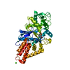 8hfnC 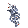 3c8zS C: citing same article ( S: Starting model for refinement |
|---|---|
| Similar structure data |
-Links
 PDBj
PDBj- Assembly
| Deposited unit | 
| ||||||||
|---|---|---|---|---|---|---|---|---|---|
| 1 |
| ||||||||
| Unit cell |
|
-Components
| #1: Protein | / L-Cys:GlcN-Ins ligase / Mycothiol ligase / MSH ligase Mass: 45508.605 Da / Num. of mol.: 1 Source method: isolated from a genetically manipulated source Source: (gene. exp.) Mycolicibacterium smegmatis MC2 155 (bacteria)Strain: ATCC 700084 / mc(2)155 / Gene: mshC, cysS2, MSMEG_4189, MSMEI_4091 / Plasmid: pETRUK / Production host: Escherichia coli (E. coli) / Strain (production host): Rosetta 2 (DE3) pLysSReferences: UniProt: A0QZY0, L-cysteine:1D-myo-inositol 2-amino-2-deoxy-alpha-D-glucopyranoside ligase |
|---|---|
| #2: Chemical | ChemComp-XG8 /   Mass: 342.457 Da / Num. of mol.: 1 / Source method: obtained synthetically / Formula: C13H14N2O3S3 / Feature type: SUBJECT OF INVESTIGATION Mass: 342.457 Da / Num. of mol.: 1 / Source method: obtained synthetically / Formula: C13H14N2O3S3 / Feature type: SUBJECT OF INVESTIGATION |
| #3: Chemical | ChemComp-ZN /   Mass: 65.409 Da / Num. of mol.: 1 / Source method: obtained synthetically / Formula: Zn Mass: 65.409 Da / Num. of mol.: 1 / Source method: obtained synthetically / Formula: Zn |
| #4: Chemical | ChemComp-CA /   Mass: 40.078 Da / Num. of mol.: 1 / Source method: obtained synthetically / Formula: Ca Mass: 40.078 Da / Num. of mol.: 1 / Source method: obtained synthetically / Formula: Ca |
| #5: Water | ChemComp-HOH / Water Mass: 18.015 Da / Num. of mol.: 10 / Source method: isolated from a natural source / Formula: H2O Mass: 18.015 Da / Num. of mol.: 10 / Source method: isolated from a natural source / Formula: H2O |
| Has ligand of interest | Y |
-Experimental details
-Experiment
| Experiment | Method: X-RAY DIFFRACTION / Number of used crystals: 1 |
|---|
- Sample preparation
| Crystal | Density Matthews: 4.69 Å3/Da / Density % sol: 73.76 % |
|---|---|
| Crystal grow | Temperature: 293 K / Method: vapor diffusion, sitting drop / pH: 8.5 Details: Holo protein at 10 mg/mL in 10 mM Tris-HCl pH 7, 100 mM NaCl and 2.5 mM 2-mercaptoethanol was mixed with 1 mM compound 7d and 1 mM TCEP (final concentrations) and incubated on ice for 1 hr. ...Details: Holo protein at 10 mg/mL in 10 mM Tris-HCl pH 7, 100 mM NaCl and 2.5 mM 2-mercaptoethanol was mixed with 1 mM compound 7d and 1 mM TCEP (final concentrations) and incubated on ice for 1 hr. Crystals of MshC in complex with compound 7d were grown in the Morpheus screen by dispensing droplets comprised of 300 nL protein mixture and 150 nL reservoir solution containing PEG550MME 20% (v/v), PEG20000 10% (w/v), Morpheus divalent 60 mM and Morpheus buffer system 3 pH 8.5. |
-Data collection
| Diffraction | Mean temperature: 100 K / Serial crystal experiment: N |
|---|---|
| Diffraction source | Source: SYNCHROTRON / Site: ESRF  / Beamline: ID23-1 / Wavelength: 0.7749 Å / Beamline: ID23-1 / Wavelength: 0.7749 Å |
| Detector | Type: DECTRIS PILATUS 6M / Detector: PIXEL / Date: Sep 24, 2021 |
| Radiation | Protocol: SINGLE WAVELENGTH / Monochromatic (M) / Laue (L): M / Scattering type: x-ray |
| Radiation wavelength | Wavelength: 0.7749 Å / Relative weight: 1 |
| Reflection | Resolution: 2.77→145.59 Å / Num. obs: 21805 / % possible obs: 100 % / Redundancy: 20.1 % / CC1/2: 0.993 / Rmerge(I) obs: 0.098 / Rpim(I) all: 0.023 / Rrim(I) all: 0.101 / Χ2: 1.04 / Net I/σ(I): 17.5 / Num. measured all: 438327 |
| Reflection shell | Resolution: 2.77→2.92 Å / % possible obs: 100 % / Redundancy: 20.6 % / Rmerge(I) obs: 1.387 / Num. measured all: 65200 / Num. unique obs: 3164 / CC1/2: 0.898 / Rpim(I) all: 0.312 / Rrim(I) all: 1.421 / Χ2: 1.01 / Net I/σ(I) obs: 2.6 |
- Processing
| Software |
| ||||||||||||||||||||||||||||||||||||||||||||||||||||||||||||||||||||||||||||||||||||||||||||||||||||||||||||||||||||||||||||||||||||||||||||||||||||||||||||||||||||||||||||||||||||||||||||||||||||||||||||||||||||||||||||||||||||||||||||||||||||||||||
|---|---|---|---|---|---|---|---|---|---|---|---|---|---|---|---|---|---|---|---|---|---|---|---|---|---|---|---|---|---|---|---|---|---|---|---|---|---|---|---|---|---|---|---|---|---|---|---|---|---|---|---|---|---|---|---|---|---|---|---|---|---|---|---|---|---|---|---|---|---|---|---|---|---|---|---|---|---|---|---|---|---|---|---|---|---|---|---|---|---|---|---|---|---|---|---|---|---|---|---|---|---|---|---|---|---|---|---|---|---|---|---|---|---|---|---|---|---|---|---|---|---|---|---|---|---|---|---|---|---|---|---|---|---|---|---|---|---|---|---|---|---|---|---|---|---|---|---|---|---|---|---|---|---|---|---|---|---|---|---|---|---|---|---|---|---|---|---|---|---|---|---|---|---|---|---|---|---|---|---|---|---|---|---|---|---|---|---|---|---|---|---|---|---|---|---|---|---|---|---|---|---|---|---|---|---|---|---|---|---|---|---|---|---|---|---|---|---|---|---|---|---|---|---|---|---|---|---|---|---|---|---|---|---|---|---|---|---|---|---|---|---|---|---|---|---|---|---|---|---|---|---|
| Refinement | Method to determine structure: MOLECULAR REPLACEMENT Starting model: 3C8Z Resolution: 2.77→55.03 Å / SU ML: 0.35 / Cross valid method: THROUGHOUT / σ(F): 1.38 / Phase error: 32.43 / Stereochemistry target values: ML
| ||||||||||||||||||||||||||||||||||||||||||||||||||||||||||||||||||||||||||||||||||||||||||||||||||||||||||||||||||||||||||||||||||||||||||||||||||||||||||||||||||||||||||||||||||||||||||||||||||||||||||||||||||||||||||||||||||||||||||||||||||||||||||
| Solvent computation | Shrinkage radii: 0.9 Å / VDW probe radii: 1.11 Å / Solvent model: FLAT BULK SOLVENT MODEL | ||||||||||||||||||||||||||||||||||||||||||||||||||||||||||||||||||||||||||||||||||||||||||||||||||||||||||||||||||||||||||||||||||||||||||||||||||||||||||||||||||||||||||||||||||||||||||||||||||||||||||||||||||||||||||||||||||||||||||||||||||||||||||
| Refinement step | Cycle: LAST / Resolution: 2.77→55.03 Å
| ||||||||||||||||||||||||||||||||||||||||||||||||||||||||||||||||||||||||||||||||||||||||||||||||||||||||||||||||||||||||||||||||||||||||||||||||||||||||||||||||||||||||||||||||||||||||||||||||||||||||||||||||||||||||||||||||||||||||||||||||||||||||||
| Refine LS restraints |
| ||||||||||||||||||||||||||||||||||||||||||||||||||||||||||||||||||||||||||||||||||||||||||||||||||||||||||||||||||||||||||||||||||||||||||||||||||||||||||||||||||||||||||||||||||||||||||||||||||||||||||||||||||||||||||||||||||||||||||||||||||||||||||
| LS refinement shell |
| ||||||||||||||||||||||||||||||||||||||||||||||||||||||||||||||||||||||||||||||||||||||||||||||||||||||||||||||||||||||||||||||||||||||||||||||||||||||||||||||||||||||||||||||||||||||||||||||||||||||||||||||||||||||||||||||||||||||||||||||||||||||||||
| Refinement TLS params. | Method: refined / Refine-ID: X-RAY DIFFRACTION
| ||||||||||||||||||||||||||||||||||||||||||||||||||||||||||||||||||||||||||||||||||||||||||||||||||||||||||||||||||||||||||||||||||||||||||||||||||||||||||||||||||||||||||||||||||||||||||||||||||||||||||||||||||||||||||||||||||||||||||||||||||||||||||
| Refinement TLS group |
|
