 Movie
Movie Controller

[English] 日本語
 Yorodumi
- PDB-8h5j: Crystal structure of PETase S121E/A180V/P181V/D186H/N233C/S242T/N... -
+ Open data

- Basic information
| Entry | Database: PDB / ID: 8h5j | ||||||
|---|---|---|---|---|---|---|---|
| Title | Crystal structure of PETase S121E/A180V/P181V/D186H/N233C/S242T/N246D/S282C mutant from Ideonella sakaiensis | ||||||
 Components Components | Poly(ethylene terephthalate) hydrolase | ||||||
 Keywords Keywords |  HYDROLASE / PET hydrolase / Ideonella sakaiensis / mutant HYDROLASE / PET hydrolase / Ideonella sakaiensis / mutant | ||||||
| Function / homology |  Function and homology information Function and homology informationpoly(ethylene terephthalate) hydrolase / acetylesterase activity / carboxylic ester hydrolase activity / : / xenobiotic catabolic process / extracellular regionSimilarity search - Function | ||||||
| Biological species |  Ideonella sakaiensis (bacteria) Ideonella sakaiensis (bacteria) | ||||||
| Method | X-RAY DIFFRACTION / SYNCHROTRON / MOLECULAR REPLACEMENT / Resolution: 1.4 Å | ||||||
 Authors Authors | Lee, S.H. / Seo, H. / Kim, K.-J. | ||||||
| Funding support | 1items
| ||||||
 Citation Citation | Journal: To Be Published Title: A case of balance engineering exhibits kinetic relationship between mesophilic and thermophilic poly(ethylene terephthalate) depolymerases. Authors: Lee, S.H. / Seo, H. / Kim, K.J. | ||||||
| History |
|
- Structure visualization
| Structure viewer | Molecule: MolmilJmol/JSmol |
|---|
- Downloads & links
-Download
| PDBx/mmCIF format | 8h5j.cif.gz | 129.9 KB | Display | PDBx/mmCIF format |
|---|---|---|---|---|
| PDB format | pdb8h5j.ent.gz | 98.9 KB | Display | PDB format |
| PDBx/mmJSON format | 8h5j.json.gz | Tree view | PDBx/mmJSON format | |
| Others |  Other downloads Other downloads |
-Validation report
| Arichive directory | https://data.pdbj.org/pub/pdb/validation_reports/h5/8h5jftp://data.pdbj.org/pub/pdb/validation_reports/h5/8h5j | HTTPS FTP |
|---|
-Related structure data
| Related structure data | 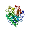 8h5kC 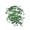 8h5lC 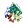 8h5mC 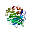 8h5oC 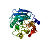 5xjhS S: Starting model for refinement C: citing same article ( |
|---|---|
| Similar structure data |
-Links
 PDBj
PDBj
- Assembly
| Deposited unit | 
| |||||||||
|---|---|---|---|---|---|---|---|---|---|---|
| 1 | 
| |||||||||
| 2 | 
| |||||||||
| Unit cell |
| |||||||||
| Components on special symmetry positions |
|
-Components
| #1: Protein | Mass: 31606.193 Da / Num. of mol.: 2 Source method: isolated from a genetically manipulated source Source: (gene. exp.) Ideonella sakaiensis (bacteria) / Gene: ISF6_4831 / Production host: Escherichia coli (E. coli)References: UniProt: A0A0K8P6T7, poly(ethylene terephthalate) hydrolase #2: Chemical |   Mass: 39.098 Da / Num. of mol.: 3 / Source method: obtained synthetically / Formula: K Mass: 39.098 Da / Num. of mol.: 3 / Source method: obtained synthetically / Formula: K#3: Chemical | Phosphate  Mass: 94.971 Da / Num. of mol.: 3 / Source method: obtained synthetically / Formula: PO4 Mass: 94.971 Da / Num. of mol.: 3 / Source method: obtained synthetically / Formula: PO4#4: Chemical | ChemComp-GOL / | Glycerol  Mass: 92.094 Da / Num. of mol.: 1 / Source method: obtained synthetically / Formula: C3H8O3 Mass: 92.094 Da / Num. of mol.: 1 / Source method: obtained synthetically / Formula: C3H8O3#5: Water | ChemComp-HOH / | Water Mass: 18.015 Da / Num. of mol.: 523 / Source method: isolated from a natural source / Formula: H2O Mass: 18.015 Da / Num. of mol.: 523 / Source method: isolated from a natural source / Formula: H2OHas ligand of interest | N | |
|---|
-Experimental details
-Experiment
| Experiment | Method: X-RAY DIFFRACTION / Number of used crystals: 1 |
|---|
- Sample preparation
| Crystal | Density Matthews: 1.86 Å3/Da / Density % sol: 33.72 % |
|---|---|
| Crystal grow | Temperature: 295 K / Method: vapor diffusion, sitting drop Details: 25 % PEG3350, 0.1 M BIS-TRIS pH6.5, and 0.2 M Ammonium sulfate |
-Data collection
| Diffraction | Mean temperature: 100 K / Serial crystal experiment: N |
|---|---|
| Diffraction source | Source: SYNCHROTRON / Site: PAL/PLS  / Beamline: 7A (6B, 6C1) / Wavelength: 0.97934 Å / Beamline: 7A (6B, 6C1) / Wavelength: 0.97934 Å |
| Detector | Type: ADSC QUANTUM 270 / Detector: CCD / Date: Mar 6, 2021 |
| Radiation | Protocol: SINGLE WAVELENGTH / Monochromatic (M) / Laue (L): M / Scattering type: x-ray |
| Radiation wavelength | Wavelength: 0.97934 Å / Relative weight: 1 |
| Reflection | Resolution: 1.4→28.65 Å / Num. obs: 88810 / % possible obs: 98.3 % / Redundancy: 1.6 % / CC1/2: 0.984 / Net I/σ(I): 30 |
| Reflection shell | Resolution: 1.4→1.42 Å / Num. unique obs: 4312 / CC1/2: 0.736 |
- Processing
| Software |
| |||||||||||||||||||||||||||||||||||||||||||||||||||||||
|---|---|---|---|---|---|---|---|---|---|---|---|---|---|---|---|---|---|---|---|---|---|---|---|---|---|---|---|---|---|---|---|---|---|---|---|---|---|---|---|---|---|---|---|---|---|---|---|---|---|---|---|---|---|---|---|---|
| Refinement | Method to determine structure: MOLECULAR REPLACEMENT Starting model: 5XJH Resolution: 1.4→28.65 Å / Cor.coef. Fo:Fc: 0.978 / Cor.coef. Fo:Fc free: 0.966 / SU B: 1.148 / SU ML: 0.044 / Cross valid method: THROUGHOUT / σ(F): 0 / ESU R: 0.06 / ESU R Free: 0.064 / Stereochemistry target values: MAXIMUM LIKELIHOOD Details: HYDROGENS HAVE BEEN ADDED IN THE RIDING POSITIONS U VALUES : REFINED INDIVIDUALLY
| |||||||||||||||||||||||||||||||||||||||||||||||||||||||
| Solvent computation | Ion probe radii: 0.8 Å / Shrinkage radii: 0.8 Å / VDW probe radii: 1.2 Å / Solvent model: MASK | |||||||||||||||||||||||||||||||||||||||||||||||||||||||
| Displacement parameters | Biso max: 77.44 Å2 / Biso mean: 17.278 Å2 / Biso min: 9.8 Å2
| |||||||||||||||||||||||||||||||||||||||||||||||||||||||
| Refinement step | Cycle: final / Resolution: 1.4→28.65 Å
| |||||||||||||||||||||||||||||||||||||||||||||||||||||||
| Refine LS restraints |
| |||||||||||||||||||||||||||||||||||||||||||||||||||||||
| LS refinement shell | Resolution: 1.404→1.441 Å / Rfactor Rfree error: 0 / Total num. of bins used: 20
|
