 Movie
Movie Controller


+ Open data

- Basic information
| Entry | Database: PDB / ID: 8h1p | ||||||||||||||||||||||||
|---|---|---|---|---|---|---|---|---|---|---|---|---|---|---|---|---|---|---|---|---|---|---|---|---|---|
| Title | Cryo-EM structure of the human RAD52 protein | ||||||||||||||||||||||||
 Components Components | DNA repair protein RAD52 homolog | ||||||||||||||||||||||||
 Keywords Keywords | RECOMBINATION / double-strand break repair / single strand annealing protein / DNA binding protein / self-oligomerization | ||||||||||||||||||||||||
| Function / homology |  Function and homology information Function and homology informationdouble-strand break repair via single-strand annealing / DNA double-strand break processing involved in repair via single-strand annealing / DNA recombinase assembly / mitotic recombination / regulation of nucleotide-excision repair / HDR through MMEJ (alt-NHEJ) / HDR through Single Strand Annealing (SSA) / SUMOylation of DNA damage response and repair proteins / protein-DNA complex / double-strand break repair via homologous recombination ...double-strand break repair via single-strand annealing / DNA double-strand break processing involved in repair via single-strand annealing / DNA recombinase assembly / mitotic recombination / regulation of nucleotide-excision repair / HDR through MMEJ (alt-NHEJ) / HDR through Single Strand Annealing (SSA) / SUMOylation of DNA damage response and repair proteins / protein-DNA complex / double-strand break repair via homologous recombination / double-strand break repair / single-stranded DNA binding / cellular response to oxidative stress / DNA recombination / protein-containing complex / DNA binding / nucleoplasm / identical protein binding / nucleusSimilarity search - Function | ||||||||||||||||||||||||
| Biological species |  Homo sapiens (human) Homo sapiens (human) | ||||||||||||||||||||||||
| Method | ELECTRON MICROSCOPY / single particle reconstruction / cryo EM / Resolution: 3.48 Å | ||||||||||||||||||||||||
 Authors Authors | Kinoshita, C. / Takizawa, Y. / Saotome, M. / Ogino, S. / Kurumizaka, H. / Kagawa, W. | ||||||||||||||||||||||||
| Funding support |  Japan, 7items Japan, 7items
| ||||||||||||||||||||||||
 Citation Citation | Journal: FEBS Open Bio / Year: 2023 Title: The cryo-EM structure of full-length RAD52 protein contains an undecameric ring. Authors: Chiaki Kinoshita / Yoshimasa Takizawa / Mika Saotome / Shun Ogino / Hitoshi Kurumizaka / Wataru Kagawa / Abstract: The human RAD52 protein, which forms an oligomeric ring structure, is involved in DNA double-strand break repair. The N-terminal half of RAD52 is primarily responsible for self-oligomerisation and ...The human RAD52 protein, which forms an oligomeric ring structure, is involved in DNA double-strand break repair. The N-terminal half of RAD52 is primarily responsible for self-oligomerisation and DNA binding. Crystallographic studies have revealed the detailed structure of the N-terminal half. However, only low-resolution structures have been reported for the full-length protein, and thus the structural role of the C-terminal half in self-oligomerisation has remained elusive. In this study, we determined the solution structure of the human RAD52 protein by cryo-electron microscopy (cryo-EM), at an average resolution of 3.5 Å. The structure revealed an undecameric ring that is nearly identical to the crystal structures of the N-terminal half. The cryo-EM map for the C-terminal half was poorly defined, indicating that the region is intrinsically disordered. The present cryo-EM structure provides important insights into the mechanistic roles played by the N-terminal and C-terminal halves of RAD52 during DNA double-strand break repair. | ||||||||||||||||||||||||
| History |
|
- Structure visualization
| Structure viewer | Molecule: MolmilJmol/JSmol |
|---|
- Downloads & links
-Download
| PDBx/mmCIF format | 8h1p.cif.gz | 415.9 KB | Display | PDBx/mmCIF format |
|---|---|---|---|---|
| PDB format | pdb8h1p.ent.gz | 329 KB | Display | PDB format |
| PDBx/mmJSON format | 8h1p.json.gz | Tree view | PDBx/mmJSON format | |
| Others |  Other downloads Other downloads |
-Validation report
| Arichive directory | https://data.pdbj.org/pub/pdb/validation_reports/h1/8h1pftp://data.pdbj.org/pub/pdb/validation_reports/h1/8h1p | HTTPS FTP |
|---|
-Related structure data
| Related structure data | 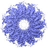 34430MC M: map data used to model this data C: citing same article ( |
|---|---|
| Similar structure data |
-Links
 PDBj
PDBj


- Assembly
| Deposited unit | 
|
|---|---|
| 1 |
|
-Components
| #1: Protein | Mass: 46514.934 Da / Num. of mol.: 11 Source method: isolated from a genetically manipulated source Source: (gene. exp.) Homo sapiens (human) / Gene: RAD52 / Production host:  Escherichia coli (E. coli) / References: UniProt: P43351 Escherichia coli (E. coli) / References: UniProt: P43351 |
|---|
-Experimental details
-Experiment
| Experiment | Method: ELECTRON MICROSCOPY |
|---|---|
| EM experiment | Aggregation state: PARTICLE / 3D reconstruction method: single particle reconstruction |
- Sample preparation
| Component | Name: RAD52 / Type: ORGANELLE OR CELLULAR COMPONENT / Entity ID: all / Source: RECOMBINANT | |||||||||||||||||||||||||
|---|---|---|---|---|---|---|---|---|---|---|---|---|---|---|---|---|---|---|---|---|---|---|---|---|---|---|
| Molecular weight | Experimental value: NO | |||||||||||||||||||||||||
| Source (natural) | Organism: Homo sapiens (human) | |||||||||||||||||||||||||
| Source (recombinant) | Organism: Escherichia coli (E. coli) | |||||||||||||||||||||||||
| Buffer solution | pH: 7.5 | |||||||||||||||||||||||||
| Buffer component |
| |||||||||||||||||||||||||
| Specimen | Conc.: 0.4 mg/ml / Embedding applied: NO / Shadowing applied: NO / Staining applied: NO / Vitrification applied: YES | |||||||||||||||||||||||||
| Specimen support | Grid material: COPPER / Grid mesh size: 200 divisions/in. / Grid type: Quantifoil R1.2/1.3 | |||||||||||||||||||||||||
| Vitrification | Instrument: FEI VITROBOT MARK IV / Cryogen name: ETHANE / Humidity: 100 % / Chamber temperature: 277 K |
- Electron microscopy imaging
| Experimental equipment |  Model: Titan Krios / Image courtesy: FEI Company |
|---|---|
| Microscopy | Model: FEI TITAN KRIOS |
| Electron gun | Electron source: FIELD EMISSION GUN / Accelerating voltage: 300 kV / Illumination mode: FLOOD BEAM |
| Electron lens | Mode: BRIGHT FIELDBright-field microscopy / Nominal defocus max: 2500 nm / Nominal defocus min: 1000 nm |
| Specimen holder | Cryogen: NITROGEN / Specimen holder model: FEI TITAN KRIOS AUTOGRID HOLDER |
| Image recording | Electron dose: 57.6 e/Å2 / Film or detector model: GATAN K3 (6k x 4k) |
- Processing
| EM software |
| ||||||||||||||||||||||||||||
|---|---|---|---|---|---|---|---|---|---|---|---|---|---|---|---|---|---|---|---|---|---|---|---|---|---|---|---|---|---|
| CTF correction | Type: PHASE FLIPPING AND AMPLITUDE CORRECTION | ||||||||||||||||||||||||||||
| 3D reconstruction | Resolution: 3.48 Å / Resolution method: FSC 0.143 CUT-OFF / Num. of particles: 39100 / Algorithm: BACK PROJECTION / Symmetry type: POINT |
