 Movie
Movie Controller


+ Open data

- Basic information
| Entry | Database: PDB / ID: 8gxo | ||||||
|---|---|---|---|---|---|---|---|
| Title | The crystal structure of CsFAOMT1 in complex with SAH | ||||||
 Components Components | Caffeoyl-CoA O-methyltransferase | ||||||
 Keywords Keywords | TRANSFERASE / CsFAOMT1 / SAH / O-methyltransferase | ||||||
| Function / homology | caffeoyl-CoA O-methyltransferase activity / lignin biosynthetic process / O-methyltransferase / Class I-like SAM-dependent O-methyltransferase / SAM-dependent O-methyltransferase class I-type profile. / methylation / S-adenosyl-L-methionine-dependent methyltransferase superfamily / S-ADENOSYL-L-HOMOCYSTEINE / Caffeoyl-CoA O-methyltransferase Function and homology information Function and homology information | ||||||
| Biological species |  Camellia sinensis (black tea) Camellia sinensis (black tea) | ||||||
| Method | X-RAY DIFFRACTION / SYNCHROTRON / MOLECULAR REPLACEMENT / Resolution: 1.74 Å | ||||||
 Authors Authors | Zhang, Z.M. / Zhou, Y.E. / Huang, H.S. | ||||||
| Funding support | 1items
| ||||||
 Citation Citation | Journal: Nat Commun / Year: 2023 Title: Characterization of two O-methyltransferases involved in the biosynthesis of O-methylated catechins in tea plant. Authors: Jin, J.Q. / Qu, F.R. / Huang, H. / Liu, Q.S. / Wei, M.Y. / Zhou, Y. / Huang, K.L. / Cui, Z. / Chen, J.D. / Dai, W.D. / Zhu, L. / Yao, M.Z. / Zhang, Z.M. / Chen, L. | ||||||
| History |
|
- Structure visualization
| Structure viewer | Molecule: MolmilJmol/JSmol |
|---|
- Downloads & links
-Download
| PDBx/mmCIF format | 8gxo.cif.gz | 138.4 KB | Display | PDBx/mmCIF format |
|---|---|---|---|---|
| PDB format | pdb8gxo.ent.gz | 85.6 KB | Display | PDB format |
| PDBx/mmJSON format | 8gxo.json.gz | Tree view | PDBx/mmJSON format | |
| Others |  Other downloads Other downloads |
-Validation report
| Arichive directory | https://data.pdbj.org/pub/pdb/validation_reports/gx/8gxoftp://data.pdbj.org/pub/pdb/validation_reports/gx/8gxo | HTTPS FTP |
|---|
-Related structure data
| Related structure data | 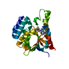 8gxnC 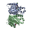 3c3yS S: Starting model for refinement C: citing same article ( |
|---|---|
| Similar structure data |
-Links
 PDBj
PDBj- Assembly
| Deposited unit | 
| ||||||||||||
|---|---|---|---|---|---|---|---|---|---|---|---|---|---|
| 1 |
| ||||||||||||
| Unit cell |
|
-Components
| #1: Protein | / CsFAOMT1 Mass: 26770.787 Da / Num. of mol.: 2 Source method: isolated from a genetically manipulated source Source: (gene. exp.) Camellia sinensis (black tea) / Gene: HYC85_015345 / Production host:  Escherichia coli (E. coli) Escherichia coli (E. coli)References: UniProt: A0A7J7GXF2, caffeoyl-CoA O-methyltransferase#2: Chemical | S-Adenosyl-L-homocysteine  Type: L-peptide linking / Mass: 384.411 Da / Num. of mol.: 2 / Source method: obtained synthetically / Formula: C14H20N6O5S / Feature type: SUBJECT OF INVESTIGATION Type: L-peptide linking / Mass: 384.411 Da / Num. of mol.: 2 / Source method: obtained synthetically / Formula: C14H20N6O5S / Feature type: SUBJECT OF INVESTIGATION#3: Chemical | ChemComp-MG / |   Mass: 24.305 Da / Num. of mol.: 1 / Source method: obtained synthetically / Formula: Mg / Feature type: SUBJECT OF INVESTIGATION Mass: 24.305 Da / Num. of mol.: 1 / Source method: obtained synthetically / Formula: Mg / Feature type: SUBJECT OF INVESTIGATION#4: Water | ChemComp-HOH / | Water Mass: 18.015 Da / Num. of mol.: 506 / Source method: isolated from a natural source / Formula: H2O Mass: 18.015 Da / Num. of mol.: 506 / Source method: isolated from a natural source / Formula: H2OHas ligand of interest | Y | |
|---|
-Experimental details
-Experiment
| Experiment | Method: X-RAY DIFFRACTION / Number of used crystals: 1 |
|---|
- Sample preparation
| Crystal | Density Matthews: 2.78 Å3/Da / Density % sol: 55.72 % |
|---|---|
| Crystal grow | Temperature: 293 K / Method: vapor diffusion, hanging drop / pH: 7.5 Details: 0.05 M Cadmium Sulfate hydrate, 0.1 M HEPES pH 7.5, 1.0 M Sodium Acetate |
-Data collection
| Diffraction | Mean temperature: 100 K / Serial crystal experiment: N |
|---|---|
| Diffraction source | Source: SYNCHROTRON / Site: CAMD  / Beamline: GCPCC / Wavelength: 0.97869 Å / Beamline: GCPCC / Wavelength: 0.97869 Å |
| Detector | Type: DECTRIS PILATUS 300K / Detector: PIXEL / Date: Dec 26, 2022 |
| Radiation | Protocol: SINGLE WAVELENGTH / Monochromatic (M) / Laue (L): M / Scattering type: x-ray |
| Radiation wavelength | Wavelength: 0.97869 Å / Relative weight: 1 |
| Reflection | Resolution: 1.74→50 Å / Num. obs: 53776 / % possible obs: 99 % / Redundancy: 20 % / Biso Wilson estimate: 26.06 Å2 / CC1/2: 1 / Net I/σ(I): 23.8 |
| Reflection shell | Resolution: 1.74→1.83 Å / Num. unique obs: 847 / CC1/2: 0.855 |
- Processing
| Software |
| |||||||||||||||||||||||||||||||||||||||||||||||||||||||||||||||||||||||||||||||||||||||||||||||||||||||||
|---|---|---|---|---|---|---|---|---|---|---|---|---|---|---|---|---|---|---|---|---|---|---|---|---|---|---|---|---|---|---|---|---|---|---|---|---|---|---|---|---|---|---|---|---|---|---|---|---|---|---|---|---|---|---|---|---|---|---|---|---|---|---|---|---|---|---|---|---|---|---|---|---|---|---|---|---|---|---|---|---|---|---|---|---|---|---|---|---|---|---|---|---|---|---|---|---|---|---|---|---|---|---|---|---|---|---|
| Refinement | Method to determine structure: MOLECULAR REPLACEMENT Starting model: 3C3Y Resolution: 1.74→43.26 Å / SU ML: 0.1844 / Cross valid method: FREE R-VALUE / σ(F): 1.35 / Phase error: 23.7012 Stereochemistry target values: GeoStd + Monomer Library + CDL v1.2
| |||||||||||||||||||||||||||||||||||||||||||||||||||||||||||||||||||||||||||||||||||||||||||||||||||||||||
| Solvent computation | Shrinkage radii: 0.9 Å / VDW probe radii: 1.1 Å / Solvent model: FLAT BULK SOLVENT MODEL | |||||||||||||||||||||||||||||||||||||||||||||||||||||||||||||||||||||||||||||||||||||||||||||||||||||||||
| Displacement parameters | Biso mean: 29.19 Å2 | |||||||||||||||||||||||||||||||||||||||||||||||||||||||||||||||||||||||||||||||||||||||||||||||||||||||||
| Refinement step | Cycle: LAST / Resolution: 1.74→43.26 Å
| |||||||||||||||||||||||||||||||||||||||||||||||||||||||||||||||||||||||||||||||||||||||||||||||||||||||||
| Refine LS restraints |
| |||||||||||||||||||||||||||||||||||||||||||||||||||||||||||||||||||||||||||||||||||||||||||||||||||||||||
| LS refinement shell |
|
