 Movie
Movie Controller


+ Open data

- Basic information
| Entry | Database: PDB / ID: 8gxn | ||||||
|---|---|---|---|---|---|---|---|
| Title | The crystal structure of CsFAOMT2 in complex with SAH | ||||||
 Components Components | caffeyl-CoA-O-methyltransferase | ||||||
 Keywords Keywords |  TRANSFERASE / CsFAOMT2 / SAH / O-methyltransferases TRANSFERASE / CsFAOMT2 / SAH / O-methyltransferases | ||||||
| Function / homology | S-ADENOSYL-L-HOMOCYSTEINE Function and homology information Function and homology information | ||||||
| Biological species |  Theaceae (tea family) Theaceae (tea family) | ||||||
| Method | X-RAY DIFFRACTION / SYNCHROTRON / MOLECULAR REPLACEMENT / Resolution: 1.34 Å | ||||||
 Authors Authors | Zhang, Z.M. / Zhou, Y.E. / Huang, H.S. | ||||||
| Funding support | 1items
| ||||||
 Citation Citation | Journal: Nat Commun / Year: 2023 Title: Characterization of two O-methyltransferases involved in the biosynthesis of O-methylated catechins in tea plant. Authors: Jin, J.Q. / Qu, F.R. / Huang, H. / Liu, Q.S. / Wei, M.Y. / Zhou, Y. / Huang, K.L. / Cui, Z. / Chen, J.D. / Dai, W.D. / Zhu, L. / Yao, M.Z. / Zhang, Z.M. / Chen, L. | ||||||
| History |
|
- Structure visualization
| Structure viewer | Molecule: MolmilJmol/JSmol |
|---|
- Downloads & links
-Download
| PDBx/mmCIF format | 8gxn.cif.gz | 130.3 KB | Display | PDBx/mmCIF format |
|---|---|---|---|---|
| PDB format | pdb8gxn.ent.gz | 81.3 KB | Display | PDB format |
| PDBx/mmJSON format | 8gxn.json.gz | Tree view | PDBx/mmJSON format | |
| Others |  Other downloads Other downloads |
-Validation report
| Arichive directory | https://data.pdbj.org/pub/pdb/validation_reports/gx/8gxnftp://data.pdbj.org/pub/pdb/validation_reports/gx/8gxn | HTTPS FTP |
|---|
-Related structure data
| Related structure data | 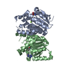 8gxoC 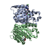 3c3yS S: Starting model for refinement C: citing same article ( |
|---|---|
| Similar structure data |
-Links
 PDBj
PDBj- Assembly
| Deposited unit | 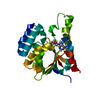
| ||||||||||||
|---|---|---|---|---|---|---|---|---|---|---|---|---|---|
| 1 |
| ||||||||||||
| 2 |
| ||||||||||||
| Unit cell |
| ||||||||||||
| Components on special symmetry positions |
|
-Components
| #1: Protein | Mass: 26628.857 Da / Num. of mol.: 1 Source method: isolated from a genetically manipulated source Source: (gene. exp.) Theaceae (tea family) / Production host:  |
|---|---|
| #2: Chemical | ChemComp-SAH / S-Adenosyl-L-homocysteine  Type: L-peptide linking / Mass: 384.411 Da / Num. of mol.: 1 / Source method: obtained synthetically / Formula: C14H20N6O5S / Feature type: SUBJECT OF INVESTIGATION Type: L-peptide linking / Mass: 384.411 Da / Num. of mol.: 1 / Source method: obtained synthetically / Formula: C14H20N6O5S / Feature type: SUBJECT OF INVESTIGATION |
| #3: Chemical | ChemComp-MG /   Mass: 24.305 Da / Num. of mol.: 1 / Source method: obtained synthetically / Formula: Mg / Feature type: SUBJECT OF INVESTIGATION Mass: 24.305 Da / Num. of mol.: 1 / Source method: obtained synthetically / Formula: Mg / Feature type: SUBJECT OF INVESTIGATION |
| #4: Water | ChemComp-HOH / Water Mass: 18.015 Da / Num. of mol.: 271 / Source method: isolated from a natural source / Formula: H2O Mass: 18.015 Da / Num. of mol.: 271 / Source method: isolated from a natural source / Formula: H2O |
| Has ligand of interest | Y |
-Experimental details
-Experiment
| Experiment | Method: X-RAY DIFFRACTION / Number of used crystals: 1 |
|---|
- Sample preparation
| Crystal | Density Matthews: 2.44 Å3/Da / Density % sol: 49.54 % |
|---|---|
| Crystal grow | Temperature: 289 K / Method: vapor diffusion, hanging drop / pH: 5.5 / Details: 1.5M Ammonium Sulfate, 100 mM Sodium Citrate pH5.5 |
-Data collection
| Diffraction | Mean temperature: 100 K / Serial crystal experiment: N |
|---|---|
| Diffraction source | Source: SYNCHROTRON / Site: CAMD  / Beamline: GCPCC / Wavelength: 0.97869 Å / Beamline: GCPCC / Wavelength: 0.97869 Å |
| Detector | Type: DECTRIS PILATUS 300K / Detector: PIXEL / Date: Oct 29, 2021 |
| Radiation | Protocol: SINGLE WAVELENGTH / Monochromatic (M) / Laue (L): M / Scattering type: x-ray |
| Radiation wavelength | Wavelength: 0.97869 Å / Relative weight: 1 |
| Reflection | Resolution: 1.28→64.74 Å / Num. obs: 61963 / % possible obs: 90.6 % / Redundancy: 15 % / Biso Wilson estimate: 15.89 Å2 / CC1/2: 1 / Net I/σ(I): 33.6 |
| Reflection shell | Resolution: 1.28→1.35 Å / Num. unique obs: 7996 / CC1/2: 0.898 |
- Processing
| Software |
| |||||||||||||||||||||||||||||||||||||||||||||||||||||||||||||||||||||||||||||||||||||||||||||||||||||||||
|---|---|---|---|---|---|---|---|---|---|---|---|---|---|---|---|---|---|---|---|---|---|---|---|---|---|---|---|---|---|---|---|---|---|---|---|---|---|---|---|---|---|---|---|---|---|---|---|---|---|---|---|---|---|---|---|---|---|---|---|---|---|---|---|---|---|---|---|---|---|---|---|---|---|---|---|---|---|---|---|---|---|---|---|---|---|---|---|---|---|---|---|---|---|---|---|---|---|---|---|---|---|---|---|---|---|---|
| Refinement | Method to determine structure: MOLECULAR REPLACEMENT Starting model: 3C3Y Resolution: 1.34→30.06 Å / SU ML: 0.1326 / Cross valid method: FREE R-VALUE / σ(F): 1.35 / Phase error: 18.5851 Stereochemistry target values: GeoStd + Monomer Library + CDL v1.2
| |||||||||||||||||||||||||||||||||||||||||||||||||||||||||||||||||||||||||||||||||||||||||||||||||||||||||
| Solvent computation | Shrinkage radii: 0.9 Å / VDW probe radii: 1.11 Å / Solvent model: FLAT BULK SOLVENT MODEL | |||||||||||||||||||||||||||||||||||||||||||||||||||||||||||||||||||||||||||||||||||||||||||||||||||||||||
| Displacement parameters | Biso mean: 22.57 Å2 | |||||||||||||||||||||||||||||||||||||||||||||||||||||||||||||||||||||||||||||||||||||||||||||||||||||||||
| Refinement step | Cycle: LAST / Resolution: 1.34→30.06 Å
| |||||||||||||||||||||||||||||||||||||||||||||||||||||||||||||||||||||||||||||||||||||||||||||||||||||||||
| Refine LS restraints |
| |||||||||||||||||||||||||||||||||||||||||||||||||||||||||||||||||||||||||||||||||||||||||||||||||||||||||
| LS refinement shell |
|
