 Movie
Movie Controller

[English] 日本語
 Yorodumi
- PDB-8fim: Structure of APOBEC3A (E72A inactive mutant) in complex with TTC-... -
+ Open data

- Basic information
| Entry | Database: PDB / ID: 8fim | ||||||
|---|---|---|---|---|---|---|---|
| Title | Structure of APOBEC3A (E72A inactive mutant) in complex with TTC-hairpin DNA substrate | ||||||
 Components Components |
| ||||||
 Keywords Keywords |  DNA BINDING PROTEIN / APOBEC / APOBEC3A / A3A / APOBEC3A E72A mutant / cancer / DNA / mutation / cytidine deaminase / hairpin DNA DNA BINDING PROTEIN / APOBEC / APOBEC3A / A3A / APOBEC3A E72A mutant / cancer / DNA / mutation / cytidine deaminase / hairpin DNA | ||||||
| Function / homology |  Function and homology information Function and homology informationmRNA Editing: C to U Conversion / Formation of the Editosome / single-stranded DNA cytosine deaminase / DNA cytosine deamination / cytidine to uridine editing / : / cytidine deaminase activity / clearance of foreign intracellular DNA / negative regulation of single stranded viral RNA replication via double stranded DNA intermediate / : ...mRNA Editing: C to U Conversion / Formation of the Editosome / single-stranded DNA cytosine deaminase / DNA cytosine deamination / cytidine to uridine editing / : / cytidine deaminase activity / clearance of foreign intracellular DNA / negative regulation of single stranded viral RNA replication via double stranded DNA intermediate / : / retrotransposon silencing / negative regulation of viral genome replication / P-body / defense response to virus / innate immune response / RNA binding / zinc ion binding / nucleoplasm / nucleus / cytoplasmSimilarity search - Function | ||||||
| Biological species |  Homo sapiens (human) Homo sapiens (human) | ||||||
| Method | X-RAY DIFFRACTION / SYNCHROTRON / MOLECULAR REPLACEMENT / Resolution: 2.22 Å | ||||||
 Authors Authors | Harjes, S. / Jameson, G.B. / Harjes, E. / Filichev, V.V. / Kurup, H.M. | ||||||
| Funding support |  New Zealand, 1items New Zealand, 1items
| ||||||
 Citation Citation | Journal: To Be Published Title: DNA-based inhibitors restrict mutagenic activity of APOBEC3 in cells Authors: Harjes, S. / Kurup, H.M. / Filichev, V. / Harjes, E. / Jameson, G.B. | ||||||
| History |
|
- Structure visualization
| Structure viewer | Molecule: MolmilJmol/JSmol |
|---|
- Downloads & links
-Download
| PDBx/mmCIF format | 8fim.cif.gz | 199.2 KB | Display | PDBx/mmCIF format |
|---|---|---|---|---|
| PDB format | pdb8fim.ent.gz | 156.9 KB | Display | PDB format |
| PDBx/mmJSON format | 8fim.json.gz | Tree view | PDBx/mmJSON format | |
| Others |  Other downloads Other downloads |
-Validation report
| Arichive directory | https://data.pdbj.org/pub/pdb/validation_reports/fi/8fimftp://data.pdbj.org/pub/pdb/validation_reports/fi/8fim | HTTPS FTP |
|---|
-Related structure data
| Related structure data | 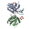 8fiiC 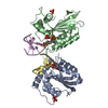 8fijC 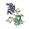 8fikC  8filC 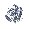 5kegS S: Starting model for refinement C: citing same article ( |
|---|---|
| Similar structure data | |
| Experimental dataset #1 | Data reference: 10.1002/cbic.201900505 / Data set type: other data |
-Links
 PDBj
PDBj


- Assembly
| Deposited unit | 
| ||||||||||||||||||||||||||||||||||||||||||||||||||||||||||||||||||||
|---|---|---|---|---|---|---|---|---|---|---|---|---|---|---|---|---|---|---|---|---|---|---|---|---|---|---|---|---|---|---|---|---|---|---|---|---|---|---|---|---|---|---|---|---|---|---|---|---|---|---|---|---|---|---|---|---|---|---|---|---|---|---|---|---|---|---|---|---|---|
| 1 |
| ||||||||||||||||||||||||||||||||||||||||||||||||||||||||||||||||||||
| 2 |
| ||||||||||||||||||||||||||||||||||||||||||||||||||||||||||||||||||||
| Unit cell |
| ||||||||||||||||||||||||||||||||||||||||||||||||||||||||||||||||||||
| Noncrystallographic symmetry (NCS) | NCS domain:
NCS domain segments: Component-ID: 0 / Refine code: 0
NCS ensembles :
|
-Components
-Protein / DNA chain , 2 types, 4 molecules ABHC
| #1: Protein | Mass: 22982.979 Da / Num. of mol.: 2 / Mutation: E72A Source method: isolated from a genetically manipulated source Source: (gene. exp.) Homo sapiens (human) / Gene: APOBEC3A / Plasmid: pETite / Production host:  Escherichia coli BL21(DE3) (bacteria) Escherichia coli BL21(DE3) (bacteria)References: UniProt: P31941, single-stranded DNA cytosine deaminase#2: DNA chain | Mass: 3934.546 Da / Num. of mol.: 2 / Source method: obtained synthetically / Source: (synth.) Homo sapiens (human) |
|---|
-Non-polymers , 5 types, 23 molecules 


| #3: Chemical | Phytic acid Mass: 660.035 Da / Num. of mol.: 2 / Source method: obtained synthetically / Formula: C6H18O24P6 Mass: 660.035 Da / Num. of mol.: 2 / Source method: obtained synthetically / Formula: C6H18O24P6#4: Chemical |  Mass: 65.409 Da / Num. of mol.: 2 / Source method: obtained synthetically / Formula: Zn Mass: 65.409 Da / Num. of mol.: 2 / Source method: obtained synthetically / Formula: Zn#5: Chemical | Chloride Mass: 35.453 Da / Num. of mol.: 2 / Source method: obtained synthetically / Formula: Cl Mass: 35.453 Da / Num. of mol.: 2 / Source method: obtained synthetically / Formula: Cl#6: Chemical | ChemComp-PO4 / | Phosphate Mass: 94.971 Da / Num. of mol.: 1 / Source method: obtained synthetically / Formula: PO4 Mass: 94.971 Da / Num. of mol.: 1 / Source method: obtained synthetically / Formula: PO4#7: Water | ChemComp-HOH / | WaterMass: 18.015 Da / Num. of mol.: 16 / Source method: isolated from a natural source / Formula: H2O |
|---|
-Details
| Has ligand of interest | N |
|---|
-Experimental details
-Experiment
| Experiment | Method: X-RAY DIFFRACTION / Number of used crystals: 1 |
|---|
- Sample preparation
| Crystal | Density Matthews: 2.51 Å3/Da / Density % sol: 56.3 % / Description: Tiny flattened needle |
|---|---|
| Crystal grow | Temperature: 285 K / Method: vapor diffusion, hanging drop / pH: 6.6 Details: A3A-E72A (50 mM MES pH 6.0, 100 mM NaCl, 1 mM TCEP, 0.2 mM EDTA) was mixed with oligonucleotides (10 mM Tris/HCl pH 7.9, 1 mM EDTA) at 0.85 mM and 1.7 mM respectively. Dilution was done with ...Details: A3A-E72A (50 mM MES pH 6.0, 100 mM NaCl, 1 mM TCEP, 0.2 mM EDTA) was mixed with oligonucleotides (10 mM Tris/HCl pH 7.9, 1 mM EDTA) at 0.85 mM and 1.7 mM respectively. Dilution was done with protein buffer. The mixture was added to crystallization liquid 1 to 1 and the mixture was pipetted on siliconized glass disks and sealed on top of a reservoir of crystallization liquid for hanging drop crystallization at 12 degrees Celsius. The crystallization liquid has the following composition: 100 mM Bicine at pH 6.6, 200 mM NaCl, 20 mM putrescine, 1 mM TCEP, 1 mM inositol hexaphosphate (phytic acid) and 45 % pentaerythritol propoxylate (5/4 PO/OH) |
-Data collection
| Diffraction | Mean temperature: 100 K / Serial crystal experiment: N |
|---|---|
| Diffraction source | Source: SYNCHROTRON / Site: Australian Synchrotron  / Beamline: MX2 / Wavelength: 0.953739 Å / Beamline: MX2 / Wavelength: 0.953739 Å |
| Detector | Type: DECTRIS EIGER X 9M / Detector: PIXEL / Date: Jun 22, 2021 / Details: 1 vertical and 2 horizontal focussing mirrors |
| Radiation | Monochromator: Si(111) double crystal monochromator / Protocol: SINGLE WAVELENGTH / Monochromatic (M) / Laue (L): M / Scattering type: x-ray |
| Radiation wavelength | Wavelength: 0.953739 Å / Relative weight: 1 |
| Reflection | Resolution: 2.22→47.99 Å / Num. obs: 26243 / % possible obs: 97.3 % / Redundancy: 3.4 % / Biso Wilson estimate: 51 Å2 / CC1/2: 0.995 / Rmerge(I) obs: 0.092 / Rpim(I) all: 0.059 / Rrim(I) all: 0.11 / Χ2: 0.46 / Net I/σ(I): 6.2 |
| Reflection shell | Resolution: 2.22→2.29 Å / Redundancy: 3.2 % / Rmerge(I) obs: 1.535 / Mean I/σ(I) obs: 0.6 / Num. unique obs: 2098 / CC1/2: 0.332 / Rpim(I) all: 0.979 / Rrim(I) all: 1.826 / Χ2: 0.31 / % possible all: 97.9 |
- Processing
| Software |
| |||||||||||||||||||||||||||||||||||||||||||||||||||||||||||||||||||||||||||||||||||||||||||||||||||||||||||||||||||||||||||||
|---|---|---|---|---|---|---|---|---|---|---|---|---|---|---|---|---|---|---|---|---|---|---|---|---|---|---|---|---|---|---|---|---|---|---|---|---|---|---|---|---|---|---|---|---|---|---|---|---|---|---|---|---|---|---|---|---|---|---|---|---|---|---|---|---|---|---|---|---|---|---|---|---|---|---|---|---|---|---|---|---|---|---|---|---|---|---|---|---|---|---|---|---|---|---|---|---|---|---|---|---|---|---|---|---|---|---|---|---|---|---|---|---|---|---|---|---|---|---|---|---|---|---|---|---|---|---|
| Refinement | Method to determine structure: MOLECULAR REPLACEMENT Starting model: 5KEG Resolution: 2.22→47.99 Å / Cor.coef. Fo:Fc: 0.937 / Cor.coef. Fo:Fc free: 0.935 / SU B: 27.009 / SU ML: 0.299 / Cross valid method: FREE R-VALUE / σ(F): 0 / ESU R: 0.322 / ESU R Free: 0.242 / Stereochemistry target values: MAXIMUM LIKELIHOOD Details: HYDROGENS HAVE BEEN ADDED IN THE RIDING POSITIONS U VALUES : WITH TLS ADDED
| |||||||||||||||||||||||||||||||||||||||||||||||||||||||||||||||||||||||||||||||||||||||||||||||||||||||||||||||||||||||||||||
| Solvent computation | Ion probe radii: 0.8 Å / Shrinkage radii: 0.8 Å / VDW probe radii: 1.2 Å / Solvent model: MASK | |||||||||||||||||||||||||||||||||||||||||||||||||||||||||||||||||||||||||||||||||||||||||||||||||||||||||||||||||||||||||||||
| Displacement parameters | Biso max: 162.28 Å2 / Biso mean: 62.63 Å2 / Biso min: 38.98 Å2
| |||||||||||||||||||||||||||||||||||||||||||||||||||||||||||||||||||||||||||||||||||||||||||||||||||||||||||||||||||||||||||||
| Refinement step | Cycle: final / Resolution: 2.22→47.99 Å
| |||||||||||||||||||||||||||||||||||||||||||||||||||||||||||||||||||||||||||||||||||||||||||||||||||||||||||||||||||||||||||||
| Refine LS restraints |
| |||||||||||||||||||||||||||||||||||||||||||||||||||||||||||||||||||||||||||||||||||||||||||||||||||||||||||||||||||||||||||||
| Refine LS restraints NCS | Refine-ID: X-RAY DIFFRACTION / Type: interatomic distance / Weight position: 0.05
| |||||||||||||||||||||||||||||||||||||||||||||||||||||||||||||||||||||||||||||||||||||||||||||||||||||||||||||||||||||||||||||
| LS refinement shell | Resolution: 2.22→2.276 Å / Rfactor Rfree error: 0
| |||||||||||||||||||||||||||||||||||||||||||||||||||||||||||||||||||||||||||||||||||||||||||||||||||||||||||||||||||||||||||||
| Refinement TLS params. | Method: refined / Refine-ID: X-RAY DIFFRACTION
| |||||||||||||||||||||||||||||||||||||||||||||||||||||||||||||||||||||||||||||||||||||||||||||||||||||||||||||||||||||||||||||
| Refinement TLS group |
|
