 Movie
Movie Controller

[English] 日本語
 Yorodumi
- PDB-8ff9: Crystal structure of Apo Dps protein (PA0962) from Pseudomonas ae... -
+ Open data

- Basic information
| Entry | Database: PDB / ID: 8ff9 | |||||||||
|---|---|---|---|---|---|---|---|---|---|---|
| Title | Crystal structure of Apo Dps protein (PA0962) from Pseudomonas aeruginosa (orthorhombic form) | |||||||||
 Components Components | Probable dna-binding stress protein | |||||||||
 Keywords Keywords | METAL BINDING PROTEIN | |||||||||
| Function / homology |  Function and homology information Function and homology informationoxidoreductase activity, acting on metal ions /  ferric iron binding / DNA binding ferric iron binding / DNA bindingSimilarity search - Function | |||||||||
| Biological species |   Pseudomonas aeruginosa (bacteria) Pseudomonas aeruginosa (bacteria) | |||||||||
| Method | X-RAY DIFFRACTION / SYNCHROTRON / MOLECULAR REPLACEMENT / Resolution: 1.7 Å | |||||||||
 Authors Authors | Lovell, S. / Kashipathy, M.M. / Battaile, K.P. / Rivera, M. | |||||||||
| Funding support |  United States, 2items United States, 2items
| |||||||||
 Citation Citation | Journal: Int J Mol Sci / Year: 2023 Title: Pseudomonas aeruginosa Dps (PA0962) Functions in H 2 O 2 Mediated Oxidative Stress Defense and Exhibits In Vitro DNA Cleaving Activity. Authors: Rajapaksha, N. / Soldano, A. / Yao, H. / Donnarumma, F. / Kashipathy, M.M. / Seibold, S. / Battaile, K.P. / Lovell, S. / Rivera, M. | |||||||||
| History |
|
- Structure visualization
| Structure viewer | Molecule: MolmilJmol/JSmol |
|---|
- Downloads & links
-Download
| PDBx/mmCIF format | 8ff9.cif.gz | 423.6 KB | Display | PDBx/mmCIF format |
|---|---|---|---|---|
| PDB format | pdb8ff9.ent.gz | 350.4 KB | Display | PDB format |
| PDBx/mmJSON format | 8ff9.json.gz | Tree view | PDBx/mmJSON format | |
| Others |  Other downloads Other downloads |
-Validation report
| Arichive directory | https://data.pdbj.org/pub/pdb/validation_reports/ff/8ff9ftp://data.pdbj.org/pub/pdb/validation_reports/ff/8ff9 | HTTPS FTP |
|---|
-Related structure data
-Links
 PDBj
PDBj


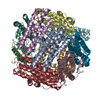
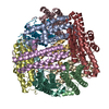
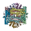
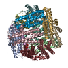
- Assembly
| Deposited unit | 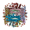
| ||||||||
|---|---|---|---|---|---|---|---|---|---|
| 1 |
| ||||||||
| Unit cell |
|
-Components
| #1: Protein | Mass: 17510.916 Da / Num. of mol.: 12 Source method: isolated from a genetically manipulated source Source: (gene. exp.) Pseudomonas aeruginosa (bacteria)Strain: ATCC 15692 / DSM 22644 / CIP 104116 / JCM 14847 / LMG 12228 / 1C / PRS 101 / PAO1 Gene: PA0962 / Plasmid: pET11a / Production host: Escherichia coli BL21(DE3) (bacteria) / Variant (production host): Gold / References: UniProt: Q9I4Z7#2: Chemical | ChemComp-CL / Chloride  Mass: 35.453 Da / Num. of mol.: 24 / Source method: obtained synthetically / Formula: Cl Mass: 35.453 Da / Num. of mol.: 24 / Source method: obtained synthetically / Formula: Cl#3: Chemical | ChemComp-SO4 / Sulfate  Mass: 96.063 Da / Num. of mol.: 81 / Source method: obtained synthetically / Formula: SO4 / Feature type: SUBJECT OF INVESTIGATION Mass: 96.063 Da / Num. of mol.: 81 / Source method: obtained synthetically / Formula: SO4 / Feature type: SUBJECT OF INVESTIGATION#4: Chemical | ChemComp-NA /   Mass: 22.990 Da / Num. of mol.: 12 / Source method: obtained synthetically / Formula: Na Mass: 22.990 Da / Num. of mol.: 12 / Source method: obtained synthetically / Formula: Na#5: Water | ChemComp-HOH / | Water Mass: 18.015 Da / Num. of mol.: 2323 / Source method: isolated from a natural source / Formula: H2O Mass: 18.015 Da / Num. of mol.: 2323 / Source method: isolated from a natural source / Formula: H2OHas ligand of interest | Y | |
|---|
-Experimental details
-Experiment
| Experiment | Method: X-RAY DIFFRACTION / Number of used crystals: 1 |
|---|
- Sample preparation
| Crystal | Density Matthews: 3.13 Å3/Da / Density % sol: 60.74 % |
|---|---|
| Crystal grow | Temperature: 291 K / Method: vapor diffusion, sitting drop / pH: 7 / Details: 1 M (NH4)2SO4, 1M KCl, 100 mM HEPES |
-Data collection
| Diffraction | Mean temperature: 291 K / Serial crystal experiment: N |
|---|---|
| Diffraction source | Source: SYNCHROTRON / Site: APS / Beamline: 17-ID / Wavelength: 1 Å |
| Detector | Type: DECTRIS EIGER2 XE 9M / Detector: PIXEL / Date: Aug 1, 2020 |
| Radiation | Monochromator: Double Crystal Si 111 / Protocol: SINGLE WAVELENGTH / Monochromatic (M) / Laue (L): M / Scattering type: x-ray |
| Radiation wavelength | Wavelength: 1 Å / Relative weight: 1 |
| Reflection | Resolution: 1.7→48.43 Å / Num. obs: 288462 / % possible obs: 100 % / Redundancy: 6.8 % / CC1/2: 0.997 / Rmerge(I) obs: 0.105 / Rpim(I) all: 0.043 / Rrim(I) all: 0.114 / Χ2: 1.02 / Net I/σ(I): 10.8 / Num. measured all: 1964310 |
| Reflection shell | Resolution: 1.7→1.73 Å / % possible obs: 100 % / Redundancy: 6.9 % / Rmerge(I) obs: 1.159 / Num. measured all: 98554 / Num. unique obs: 14185 / CC1/2: 0.632 / Rpim(I) all: 0.469 / Rrim(I) all: 1.252 / Χ2: 1.06 / Net I/σ(I) obs: 1.8 |
- Processing
| Software |
| |||||||||||||||||||||||||||||||||||||||||||||||||||||||||||||||||||||||||||||||||||||||||||||||||||||||||||||||||||||||||||||||||||||||||||||||||||||||||||||||||||||||||||||||||||||||||||||||||||||||||||||||||||||||||
|---|---|---|---|---|---|---|---|---|---|---|---|---|---|---|---|---|---|---|---|---|---|---|---|---|---|---|---|---|---|---|---|---|---|---|---|---|---|---|---|---|---|---|---|---|---|---|---|---|---|---|---|---|---|---|---|---|---|---|---|---|---|---|---|---|---|---|---|---|---|---|---|---|---|---|---|---|---|---|---|---|---|---|---|---|---|---|---|---|---|---|---|---|---|---|---|---|---|---|---|---|---|---|---|---|---|---|---|---|---|---|---|---|---|---|---|---|---|---|---|---|---|---|---|---|---|---|---|---|---|---|---|---|---|---|---|---|---|---|---|---|---|---|---|---|---|---|---|---|---|---|---|---|---|---|---|---|---|---|---|---|---|---|---|---|---|---|---|---|---|---|---|---|---|---|---|---|---|---|---|---|---|---|---|---|---|---|---|---|---|---|---|---|---|---|---|---|---|---|---|---|---|---|---|---|---|---|---|---|---|---|---|---|---|---|---|---|---|---|
| Refinement | Method to determine structure: MOLECULAR REPLACEMENT / Resolution: 1.7→48.43 Å / SU ML: 0.16 / Cross valid method: FREE R-VALUE / σ(F): 1.01 / Phase error: 16.17 / Stereochemistry target values: ML
| |||||||||||||||||||||||||||||||||||||||||||||||||||||||||||||||||||||||||||||||||||||||||||||||||||||||||||||||||||||||||||||||||||||||||||||||||||||||||||||||||||||||||||||||||||||||||||||||||||||||||||||||||||||||||
| Solvent computation | Shrinkage radii: 0.9 Å / VDW probe radii: 1.11 Å / Solvent model: FLAT BULK SOLVENT MODEL | |||||||||||||||||||||||||||||||||||||||||||||||||||||||||||||||||||||||||||||||||||||||||||||||||||||||||||||||||||||||||||||||||||||||||||||||||||||||||||||||||||||||||||||||||||||||||||||||||||||||||||||||||||||||||
| Refinement step | Cycle: LAST / Resolution: 1.7→48.43 Å
| |||||||||||||||||||||||||||||||||||||||||||||||||||||||||||||||||||||||||||||||||||||||||||||||||||||||||||||||||||||||||||||||||||||||||||||||||||||||||||||||||||||||||||||||||||||||||||||||||||||||||||||||||||||||||
| Refine LS restraints |
| |||||||||||||||||||||||||||||||||||||||||||||||||||||||||||||||||||||||||||||||||||||||||||||||||||||||||||||||||||||||||||||||||||||||||||||||||||||||||||||||||||||||||||||||||||||||||||||||||||||||||||||||||||||||||
| LS refinement shell |
|
