Movie
Movie Controller


+ Open data

- Basic information
| Entry | Database: PDB / ID: 8fcg | ||||||
|---|---|---|---|---|---|---|---|
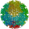
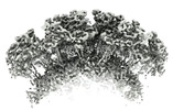
| Title | Cryo-EM structure of Chikungunya virus asymmetric unit | ||||||
 Components Components |
| ||||||
 Keywords Keywords |  VIRUS / CHIKV asymmetric unit VIRUS / CHIKV asymmetric unit | ||||||
| Function / homology |  Function and homology information Function and homology informationT=4 icosahedral viral capsid / host cell cytoplasm / symbiont entry into host cell / serine-type endopeptidase activity / fusion of virus membrane with host endosome membrane / virion attachment to host cell / host cell plasma membrane / virion membrane / structural molecule activity / proteolysis ...T=4 icosahedral viral capsid / host cell cytoplasm / symbiont entry into host cell / serine-type endopeptidase activity / fusion of virus membrane with host endosome membrane / virion attachment to host cell / host cell plasma membrane / virion membrane / structural molecule activity / proteolysis / plasma membrane / cytoplasmSimilarity search - Function | ||||||
| Biological species |   Chikungunya virus Chikungunya virus | ||||||
| Method | ELECTRON MICROSCOPY / single particle reconstruction / cryo EM / Resolution: 3.09 Å | ||||||
 Authors Authors | Su, G.C. / Chmielewsk, D. / Kaelber, J. / Pintilie, G. / Chen, M. / Jin, J. / Auguste, A. / Chiu, W. | ||||||
| Funding support |  United States, 1items United States, 1items
| ||||||
 Citation Citation | Journal: PNAS Nexus / Year: 2024 Title: Cryogenic electron microscopy and tomography reveal imperfect icosahedral symmetry in alphaviruses. Authors: David Chmielewski / Guan-Chin Su / Jason T Kaelber / Grigore D Pintilie / Muyuan Chen / Jing Jin / Albert J Auguste / Wah Chiu / Abstract: Alphaviruses are spherical, enveloped RNA viruses with two-layered icosahedral architecture. The structures of many alphaviruses have been studied using cryogenic electron microscopy (cryo-EM) ...Alphaviruses are spherical, enveloped RNA viruses with two-layered icosahedral architecture. The structures of many alphaviruses have been studied using cryogenic electron microscopy (cryo-EM) reconstructions, which impose icosahedral symmetry on the viral particles. Using cryogenic electron tomography (cryo-ET), we revealed a polarized symmetry defect in the icosahedral lattice of Chikungunya virus (CHIKV) in situ, similar to the late budding particles, suggesting the inherent imperfect symmetry originates from the final pinch-off of assembled virions. We further demonstrated this imperfect symmetry is also present in in vitro purified CHIKV and Mayaro virus, another arthritogenic alphavirus. We employed a subparticle-based single-particle analysis protocol to circumvent the icosahedral imperfection and boosted the resolution of the structure of the CHIKV to ∼3 Å resolution, which revealed detailed molecular interactions between glycoprotein E1-E2 heterodimers in the transmembrane region and multiple lipid-like pocket factors located in a highly conserved hydrophobic pocket. This complementary use of in situ cryo-ET and single-particle cryo-EM approaches provides a more precise structural description of near-icosahedral viruses and valuable insights to guide the development of structure-based antiviral therapies against alphaviruses. | ||||||
| History |
|
- Structure visualization
| Structure viewer | Molecule: MolmilJmol/JSmol |
|---|
- Downloads & links
-Download
| PDBx/mmCIF format | 8fcg.cif.gz | 759.4 KB | Display | PDBx/mmCIF format |
|---|---|---|---|---|
| PDB format | pdb8fcg.ent.gz | Display | PDB format | |
| PDBx/mmJSON format | 8fcg.json.gz | Tree view | PDBx/mmJSON format | |
| Others |  Other downloads Other downloads |
-Validation report
| Arichive directory | https://data.pdbj.org/pub/pdb/validation_reports/fc/8fcgftp://data.pdbj.org/pub/pdb/validation_reports/fc/8fcg | HTTPS FTP |
|---|
-Related structure data
| Related structure data |  28979MC M: map data used to model this data C: citing same article ( |
|---|---|
| Similar structure data |


-Links
 PDBj
PDBj


- Assembly
| Deposited unit | 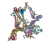
|
|---|---|
| 1 |
|
-Components
| #1: Protein | Mass: 47497.906 Da / Num. of mol.: 4 / Fragment: UNP residues 810-1248 / Source method: isolated from a natural source / Source: (natural) Chikungunya virus / Strain: vaccine strain 181/clone 25 / References: UniProt: Q88628, togavirin#2: Protein | Mass: 47077.719 Da / Num. of mol.: 4 / Fragment: UNP residues 330-748 / Source method: isolated from a natural source / Source: (natural) Chikungunya virus / Strain: vaccine strain 181/clone 25 / References: UniProt: Q88628, togavirin#3: Protein | CapsidMass: 16428.607 Da / Num. of mol.: 4 / Fragment: UNP residues 111-261 / Source method: isolated from a natural source / Source: (natural) Chikungunya virus / Strain: vaccine strain 181/clone 25 / References: UniProt: Q88628, togavirin#4: Chemical | ChemComp-PCW /   Mass: 787.121 Da / Num. of mol.: 5 Mass: 787.121 Da / Num. of mol.: 5Source method: isolated from a genetically manipulated source Formula: C44H85NO8P / Feature type: SUBJECT OF INVESTIGATION / Comment: DOPC, phospholipid*YM Has ligand of interest | Y | |
|---|
-Experimental details
-Experiment
| Experiment | Method: ELECTRON MICROSCOPY |
|---|---|
| EM experiment | Aggregation state: PARTICLE / 3D reconstruction method: single particle reconstruction |
- Sample preparation
| Component | Name: Chikungunya virusChikungunya / Type: VIRUS / Entity ID: #1-#3 / Source: NATURAL | ||||||||||||||||
|---|---|---|---|---|---|---|---|---|---|---|---|---|---|---|---|---|---|
| Molecular weight | Value: 0.5 MDa / Experimental value: NO | ||||||||||||||||
| Source (natural) | Organism: Chikungunya virus / Strain: vaccine strain 181/clone 25 | ||||||||||||||||
| Details of virus | Empty: NO / Enveloped: YES / Isolate: STRAIN / Type: VIRION | ||||||||||||||||
| Virus shell | Diameter: 700 nm / Triangulation number (T number): 4 | ||||||||||||||||
| Buffer solution | pH: 8 | ||||||||||||||||
| Buffer component |
| ||||||||||||||||
| Specimen | Embedding applied: NO / Shadowing applied: NO / Staining applied: NO / Vitrification applied: YES | ||||||||||||||||
| Specimen support | Grid material: COPPER / Grid mesh size: 200 divisions/in. / Grid type: Quantifoil R2/1 | ||||||||||||||||
| Vitrification | Instrument: LEICA EM GP / Cryogen name: ETHANE / Humidity: 100 % / Chamber temperature: 293.15 K |
- Electron microscopy imaging
| Experimental equipment |  Model: Titan Krios / Image courtesy: FEI Company |
|---|---|
| Microscopy | Model: FEI TITAN KRIOS |
| Electron gun | Electron source: FIELD EMISSION GUN / Accelerating voltage: 300 kV / Illumination mode: FLOOD BEAM |
| Electron lens | Mode: BRIGHT FIELDBright-field microscopy / Nominal magnification: 106000 X / Nominal defocus max: 2400 nm / Nominal defocus min: 1000 nm / Cs: 2.7 mm / C2 aperture diameter: 70 µm / Alignment procedure: COMA FREE |
| Specimen holder | Cryogen: NITROGEN / Specimen holder model: FEI TITAN KRIOS AUTOGRID HOLDER |
| Image recording | Electron dose: 47.25 e/Å2 / Detector mode: COUNTING / Film or detector model: GATAN K2 SUMMIT (4k x 4k) |
| EM imaging optics | Energyfilter name: GIF Bioquantum / Energyfilter slit width: 20 eV |
| Image scans | Movie frames/image: 40 |
- Processing
| EM software |
| ||||||||||||||||||||||||||||||||||||||||||||
|---|---|---|---|---|---|---|---|---|---|---|---|---|---|---|---|---|---|---|---|---|---|---|---|---|---|---|---|---|---|---|---|---|---|---|---|---|---|---|---|---|---|---|---|---|---|
| CTF correction | Type: PHASE FLIPPING AND AMPLITUDE CORRECTION | ||||||||||||||||||||||||||||||||||||||||||||
| Particle selection | Num. of particles selected: 142609 | ||||||||||||||||||||||||||||||||||||||||||||
| Symmetry | Point symmetry: C1 (asymmetric) | ||||||||||||||||||||||||||||||||||||||||||||
| 3D reconstruction | Resolution: 3.09 Å / Resolution method: FSC 0.143 CUT-OFF / Num. of particles: 2252628 / Algorithm: FOURIER SPACE / Num. of class averages: 2 / Symmetry type: POINT | ||||||||||||||||||||||||||||||||||||||||||||
| Atomic model building | Protocol: FLEXIBLE FIT / Space: REAL | ||||||||||||||||||||||||||||||||||||||||||||
| Atomic model building | PDB-ID: 6NK5 Accession code: 6NK5 / Source name: PDB / Type: experimental model | ||||||||||||||||||||||||||||||||||||||||||||
| Refine LS restraints |
|