 Movie
Movie Controller

[English] 日本語
 Yorodumi
- PDB-8ei6: Crystal structure of the WWP2 HECT domain in complex with H305, a... -
+ Open data

- Basic information
| Entry | Database: PDB / ID: 8ei6 | ||||||
|---|---|---|---|---|---|---|---|
| Title | Crystal structure of the WWP2 HECT domain in complex with H305, a Helicon Polypeptide | ||||||
 Components Components |
| ||||||
 Keywords Keywords |  LIGASE / E3 ligase / complex / stapled peptide LIGASE / E3 ligase / complex / stapled peptide | ||||||
| Function / homology |  Function and homology information Function and homology informationnegative regulation of protein transport / extracellular transport / regulation of potassium ion transmembrane transporter activity / HECT-type E3 ubiquitin transferase / negative regulation of transporter activity / regulation of monoatomic ion transmembrane transport / negative regulation of Notch signaling pathway / RHOJ GTPase cycle / RHOQ GTPase cycle / protein K63-linked ubiquitination ...negative regulation of protein transport / extracellular transport / regulation of potassium ion transmembrane transporter activity / HECT-type E3 ubiquitin transferase / negative regulation of transporter activity / regulation of monoatomic ion transmembrane transport / negative regulation of Notch signaling pathway / RHOJ GTPase cycle / RHOQ GTPase cycle / protein K63-linked ubiquitination / RHOU GTPase cycle / transcription factor binding / protein autoubiquitination / ubiquitin ligase complex / regulation of membrane potential / NOTCH3 Activation and Transmission of Signal to the Nucleus / protein modification process / negative regulation of DNA-binding transcription factor activity / Regulation of PTEN stability and activity / ubiquitin-protein transferase activity / ubiquitin protein ligase activity / proteasome-mediated ubiquitin-dependent protein catabolic process / RNA polymerase II-specific DNA-binding transcription factor binding / transcription by RNA polymerase II / protein ubiquitination / symbiont entry into host cell / negative regulation of gene expression / negative regulation of DNA-templated transcription / negative regulation of transcription by RNA polymerase II / positive regulation of transcription by RNA polymerase II / extracellular exosome / membrane / nucleus / cytosol / cytoplasmSimilarity search - Function | ||||||
| Biological species |  Homo sapiens (human) Homo sapiens (human)synthetic construct (others) | ||||||
| Method | X-RAY DIFFRACTION / SYNCHROTRON / MOLECULAR REPLACEMENT / Resolution: 3.62 Å | ||||||
 Authors Authors | Li, K. / Tokareva, O.S. / Thomson, T.M. / Verdine, G.L. / McGee, J.H. | ||||||
| Funding support |  United States, 1items United States, 1items
| ||||||
 Citation Citation | Journal: Nat Commun / Year: 2023 Title: Recognition and reprogramming of E3 ubiquitin ligase surfaces by alpha-helical peptides. Authors: Tokareva, O.S. / Li, K. / Travaline, T.L. / Thomson, T.M. / Swiecicki, J.M. / Moussa, M. / Ramirez, J.D. / Litchman, S. / Verdine, G.L. / McGee, J.H. | ||||||
| History |
|
- Structure visualization
| Structure viewer | Molecule: MolmilJmol/JSmol |
|---|
- Downloads & links
-Download
| PDBx/mmCIF format | 8ei6.cif.gz | 174.3 KB | Display | PDBx/mmCIF format |
|---|---|---|---|---|
| PDB format | pdb8ei6.ent.gz | 136.8 KB | Display | PDB format |
| PDBx/mmJSON format | 8ei6.json.gz | Tree view | PDBx/mmJSON format | |
| Others |  Other downloads Other downloads |
-Validation report
| Arichive directory | https://data.pdbj.org/pub/pdb/validation_reports/ei/8ei6ftp://data.pdbj.org/pub/pdb/validation_reports/ei/8ei6 | HTTPS FTP |
|---|
-Related structure data
| Related structure data | 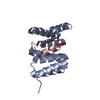 8ehzC  8ei0C 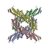 8ei1C 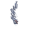 8ei2C  8ei3C  8ei4C 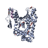 8ei5C 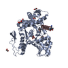 8ei7C 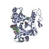 8ei8C 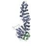 8ei9C 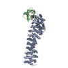 8eiaC  8eibC 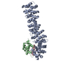 8eicC  4y07S S: Starting model for refinement C: citing same article ( |
|---|---|
| Similar structure data |
-Links
 PDBj
PDBj


- Assembly
| Deposited unit | 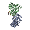
| |||||||||||||||||||||||||||||||||||||||||||||||||||||||||||||||||||||||||||||||||||
|---|---|---|---|---|---|---|---|---|---|---|---|---|---|---|---|---|---|---|---|---|---|---|---|---|---|---|---|---|---|---|---|---|---|---|---|---|---|---|---|---|---|---|---|---|---|---|---|---|---|---|---|---|---|---|---|---|---|---|---|---|---|---|---|---|---|---|---|---|---|---|---|---|---|---|---|---|---|---|---|---|---|---|---|---|
| 1 | 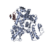
| |||||||||||||||||||||||||||||||||||||||||||||||||||||||||||||||||||||||||||||||||||
| 2 | 
| |||||||||||||||||||||||||||||||||||||||||||||||||||||||||||||||||||||||||||||||||||
| Unit cell |
| |||||||||||||||||||||||||||||||||||||||||||||||||||||||||||||||||||||||||||||||||||
| Noncrystallographic symmetry (NCS) | NCS domain:
NCS domain segments:
|