 Movie
Movie Controller

[English] 日本語
 Yorodumi
- PDB-8ehz: Crystal structure of the STUB1 TPR domain in complex with H317, a... -
+ Open data

- Basic information
| Entry | Database: PDB / ID: 8ehz | ||||||
|---|---|---|---|---|---|---|---|
| Title | Crystal structure of the STUB1 TPR domain in complex with H317, a Helicon Polypeptide | ||||||
 Components Components |
| ||||||
 Keywords Keywords |  LIGASE / E3 ligase / complex / stapled peptide LIGASE / E3 ligase / complex / stapled peptide | ||||||
| Function / homology |  Function and homology information Function and homology informationpositive regulation of chaperone-mediated protein complex assembly / regulation of glucocorticoid metabolic process / ubiquitin conjugating enzyme complex / positive regulation of ERAD pathway / positive regulation of mitophagy / ERBB2 signaling pathway / positive regulation of smooth muscle cell apoptotic process / nuclear inclusion body / misfolded protein binding / cellular response to misfolded protein ...positive regulation of chaperone-mediated protein complex assembly / regulation of glucocorticoid metabolic process / ubiquitin conjugating enzyme complex / positive regulation of ERAD pathway / positive regulation of mitophagy / ERBB2 signaling pathway / positive regulation of smooth muscle cell apoptotic process / nuclear inclusion body / misfolded protein binding / cellular response to misfolded protein / protein folding chaperone complex / ERAD pathway / ubiquitin-ubiquitin ligase activity / RIPK1-mediated regulated necrosis / chaperone-mediated autophagy / protein quality control for misfolded or incompletely synthesized proteins / TPR domain binding / protein monoubiquitination / SMAD binding / negative regulation of smooth muscle cell apoptotic process / protein K63-linked ubiquitination / positive regulation of proteolysis / R-SMAD binding / protein maturation / protein autoubiquitination / ubiquitin ligase complex / endoplasmic reticulum unfolded protein response / Hsp70 protein binding / heat shock protein binding / Downregulation of TGF-beta receptor signaling / positive regulation of protein ubiquitination / response to ischemia / G protein-coupled receptor binding / Regulation of TNFR1 signaling / negative regulation of transforming growth factor beta receptor signaling pathway / Hsp90 protein binding / regulation of protein stability / RING-type E3 ubiquitin transferase / tau protein binding / Regulation of necroptotic cell death / Downregulation of ERBB2 signaling / Z disc / kinase binding / Regulation of PTEN stability and activity / protein polyubiquitination / ubiquitin-protein transferase activity / Regulation of RUNX2 expression and activity / ubiquitin protein ligase activity / MAPK cascade / protein-macromolecule adaptor activity / Antigen processing: Ubiquitination & Proteasome degradation / positive regulation of proteasomal ubiquitin-dependent protein catabolic process / cellular response to heat / ubiquitin-dependent protein catabolic process / cellular response to hypoxia / protein-folding chaperone binding / proteasome-mediated ubiquitin-dependent protein catabolic process / protein stabilization / protein ubiquitination / DNA repair / ubiquitin protein ligase binding / enzyme binding / endoplasmic reticulum / protein homodimerization activity / mitochondrion / nucleoplasm / nucleus / cytosol / cytoplasmSimilarity search - Function | ||||||
| Biological species |  Homo sapiens (human) Homo sapiens (human)synthetic construct (others) | ||||||
| Method | X-RAY DIFFRACTION / SYNCHROTRON / MOLECULAR REPLACEMENT / Resolution: 2.06 Å | ||||||
 Authors Authors | Li, K. / Swiecicki, J.-M. / Tokareva, O.S. / Thomson, T.M. / Verdine, G.L. / McGee, J.H. | ||||||
| Funding support |  United States, 1items United States, 1items
| ||||||
 Citation Citation | Journal: Nat Commun / Year: 2023 Title: Recognition and reprogramming of E3 ubiquitin ligase surfaces by alpha-helical peptides. Authors: Tokareva, O.S. / Li, K. / Travaline, T.L. / Thomson, T.M. / Swiecicki, J.M. / Moussa, M. / Ramirez, J.D. / Litchman, S. / Verdine, G.L. / McGee, J.H. | ||||||
| History |
|
- Structure visualization
| Structure viewer | Molecule: MolmilJmol/JSmol |
|---|
- Downloads & links
-Download
| PDBx/mmCIF format | 8ehz.cif.gz | 71.5 KB | Display | PDBx/mmCIF format |
|---|---|---|---|---|
| PDB format | pdb8ehz.ent.gz | 50.8 KB | Display | PDB format |
| PDBx/mmJSON format | 8ehz.json.gz | Tree view | PDBx/mmJSON format | |
| Others |  Other downloads Other downloads |
-Validation report
| Arichive directory | https://data.pdbj.org/pub/pdb/validation_reports/eh/8ehzftp://data.pdbj.org/pub/pdb/validation_reports/eh/8ehz | HTTPS FTP |
|---|
-Related structure data
| Related structure data |  8ei0C 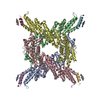 8ei1C 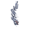 8ei2C  8ei3C  8ei4C 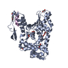 8ei5C 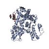 8ei6C  8ei7C  8ei8C  8ei9C  8eiaC  8eibC 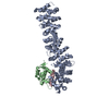 8eicC 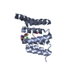 6nsvS S: Starting model for refinement C: citing same article ( |
|---|---|
| Similar structure data |
-Links
 PDBj
PDBj


- Assembly
| Deposited unit | 
| ||||||||||
|---|---|---|---|---|---|---|---|---|---|---|---|
| 1 | 
| ||||||||||
| 2 | 
| ||||||||||
| Unit cell |
|
-Components
| #1: Protein/peptide | Mass: 1277.491 Da / Num. of mol.: 2 / Source method: obtained synthetically / Source: (synth.) synthetic construct (others) #2: Protein | Mass: 15884.057 Da / Num. of mol.: 2 / Fragment: TPR domain Source method: isolated from a genetically manipulated source Source: (gene. exp.) Homo sapiens (human) / Gene: STUB1, CHIP, PP1131 / Production host:  Escherichia coli (E. coli) Escherichia coli (E. coli)References: UniProt: Q9UNE7, RING-type E3 ubiquitin transferase #3: Chemical |   Mass: 192.214 Da / Num. of mol.: 2 / Source method: obtained synthetically / Formula: C10H12N2O2 Mass: 192.214 Da / Num. of mol.: 2 / Source method: obtained synthetically / Formula: C10H12N2O2#4: Water | ChemComp-HOH / | Water Mass: 18.015 Da / Num. of mol.: 18 / Source method: isolated from a natural source / Formula: H2O Mass: 18.015 Da / Num. of mol.: 18 / Source method: isolated from a natural source / Formula: H2OHas ligand of interest | N | |
|---|
-Experimental details
-Experiment
| Experiment | Method: X-RAY DIFFRACTION / Number of used crystals: 1 |
|---|
- Sample preparation
| Crystal | Density Matthews: 2.35 Å3/Da / Density % sol: 47.57 % |
|---|---|
| Crystal grow | Temperature: 291 K / Method: vapor diffusion, hanging drop / pH: 6 Details: 0.1M Sodium acetate, 0.1M sodium citrate pH6.0, 10% PEG4000 |
-Data collection
| Diffraction | Mean temperature: 100 K / Serial crystal experiment: N |
|---|---|
| Diffraction source | Source: SYNCHROTRON / Site: APS / Beamline: 23-ID-D / Wavelength: 1.033 Å |
| Detector | Type: DECTRIS PILATUS3 6M / Detector: PIXEL / Date: Nov 6, 2020 |
| Radiation | Protocol: SINGLE WAVELENGTH / Monochromatic (M) / Laue (L): M / Scattering type: x-ray |
| Radiation wavelength | Wavelength: 1.033 Å / Relative weight: 1 |
| Reflection | Resolution: 2.06→43.08 Å / Num. obs: 19293 / % possible obs: 100 % / Redundancy: 10.5 % / Biso Wilson estimate: 44.97 Å2 / CC1/2: 0.998 / Rmerge(I) obs: 0.078 / Net I/σ(I): 15.2 |
| Reflection shell | Resolution: 2.06→2.12 Å / Redundancy: 10.6 % / Rmerge(I) obs: 0.796 / Mean I/σ(I) obs: 2.6 / Num. unique obs: 1471 / CC1/2: 0.938 / % possible all: 100 |
- Processing
| Software |
| |||||||||||||||||||||||||||||||||||||||||||||||||||||||||||||||||||||||||||||||||||||||||||||||||||||||||
|---|---|---|---|---|---|---|---|---|---|---|---|---|---|---|---|---|---|---|---|---|---|---|---|---|---|---|---|---|---|---|---|---|---|---|---|---|---|---|---|---|---|---|---|---|---|---|---|---|---|---|---|---|---|---|---|---|---|---|---|---|---|---|---|---|---|---|---|---|---|---|---|---|---|---|---|---|---|---|---|---|---|---|---|---|---|---|---|---|---|---|---|---|---|---|---|---|---|---|---|---|---|---|---|---|---|---|
| Refinement | Method to determine structure: MOLECULAR REPLACEMENT Starting model: 6NSV Resolution: 2.06→43.08 Å / SU ML: 0.3 / Cross valid method: THROUGHOUT / σ(F): 1.99 / Phase error: 39.04 / Stereochemistry target values: ML
| |||||||||||||||||||||||||||||||||||||||||||||||||||||||||||||||||||||||||||||||||||||||||||||||||||||||||
| Solvent computation | Shrinkage radii: 0.9 Å / VDW probe radii: 1.11 Å / Solvent model: FLAT BULK SOLVENT MODEL | |||||||||||||||||||||||||||||||||||||||||||||||||||||||||||||||||||||||||||||||||||||||||||||||||||||||||
| Displacement parameters | Biso max: 99.22 Å2 / Biso mean: 60.7906 Å2 / Biso min: 29.48 Å2 | |||||||||||||||||||||||||||||||||||||||||||||||||||||||||||||||||||||||||||||||||||||||||||||||||||||||||
| Refinement step | Cycle: final / Resolution: 2.06→43.08 Å
| |||||||||||||||||||||||||||||||||||||||||||||||||||||||||||||||||||||||||||||||||||||||||||||||||||||||||
| LS refinement shell | Refine-ID: X-RAY DIFFRACTION / Rfactor Rfree error: 0 / Total num. of bins used: 14
|
