 Movie
Movie Controller


+ Open data

- Basic information
| Entry | Database: PDB / ID: 8efu | ||||||
|---|---|---|---|---|---|---|---|
| Title | a22L prion fibril | ||||||
 Components Components | Major prion protein | ||||||
 Keywords Keywords | PROTEIN FIBRIL /  Prion / Fibril / GPI-anchorless / PrP / Infectious / Amyloid / Brain-derived / ex vivo / Prion strain Prion / Fibril / GPI-anchorless / PrP / Infectious / Amyloid / Brain-derived / ex vivo / Prion strain | ||||||
| Function / homology |  Function and homology information Function and homology informationInsertion of tail-anchored proteins into the endoplasmic reticulum membrane / negative regulation of amyloid precursor protein catabolic process / lamin binding / regulation of glutamate receptor signaling pathway / regulation of calcium ion import across plasma membrane / aspartic-type endopeptidase inhibitor activity / glycosaminoglycan binding / negative regulation of interleukin-17 production / ATP-dependent protein binding / regulation of potassium ion transmembrane transport ...Insertion of tail-anchored proteins into the endoplasmic reticulum membrane / negative regulation of amyloid precursor protein catabolic process / lamin binding / regulation of glutamate receptor signaling pathway / regulation of calcium ion import across plasma membrane / aspartic-type endopeptidase inhibitor activity / glycosaminoglycan binding / negative regulation of interleukin-17 production / ATP-dependent protein binding / regulation of potassium ion transmembrane transport / negative regulation of dendritic spine maintenance / type 5 metabotropic glutamate receptor binding / cupric ion binding / nucleobase-containing compound metabolic process / response to copper ion / negative regulation of calcineurin-NFAT signaling cascade / negative regulation of interleukin-2 production / negative regulation of T cell receptor signaling pathway / cuprous ion binding / activation of protein kinase activity / negative regulation of amyloid-beta formation / negative regulation of activated T cell proliferation / response to amyloid-beta / : / negative regulation of type II interferon production / intracellular copper ion homeostasis / positive regulation of protein targeting to membrane / negative regulation of long-term synaptic potentiation / response to cadmium ion / side of membrane / inclusion body / regulation of peptidyl-tyrosine phosphorylation / cellular response to copper ion / neuron projection maintenance / molecular condensate scaffold activity / tubulin binding / protein sequestering activity / negative regulation of protein phosphorylation / molecular function activator activity / positive regulation of protein localization to plasma membrane / protein destabilization / protein homooligomerization / negative regulation of DNA-binding transcription factor activity / terminal bouton / cellular response to amyloid-beta / positive regulation of neuron apoptotic process / regulation of protein localization / positive regulation of peptidyl-tyrosine phosphorylation / cellular response to xenobiotic stimulus / signaling receptor activity / amyloid-beta binding / protein-folding chaperone binding / microtubule binding / nuclear membrane / response to oxidative stress / protease binding / mitochondrial outer membrane / transmembrane transporter binding / postsynaptic density / learning or memory / molecular adaptor activity / membrane raft / copper ion binding / intracellular membrane-bounded organelle / dendrite / protein-containing complex binding / negative regulation of apoptotic process / Golgi apparatus / cell surface / endoplasmic reticulum / membrane / identical protein binding / metal ion binding / plasma membrane / cytosolSimilarity search - Function | ||||||
| Biological species |  Mus musculus (house mouse) Mus musculus (house mouse) | ||||||
| Method | ELECTRON MICROSCOPY / helical reconstruction / cryo EM / Resolution: 3.2 Å | ||||||
 Authors Authors | Hoyt, F. / Caughey, B. | ||||||
| Funding support |  United States, 1items United States, 1items
| ||||||
 Citation Citation | Journal: PLoS Pathog / Year: 2022 Title: Cryo-EM of prion strains from the same genotype of host identifies conformational determinants. Authors: Forrest Hoyt / Parvez Alam / Efrosini Artikis / Cindi L Schwartz / Andrew G Hughson / Brent Race / Chase Baune / Gregory J Raymond / Gerald S Baron / Allison Kraus / Byron Caughey / Abstract: Prion strains in a given type of mammalian host are distinguished by differences in clinical presentation, neuropathological lesions, survival time, and characteristics of the infecting prion protein ...Prion strains in a given type of mammalian host are distinguished by differences in clinical presentation, neuropathological lesions, survival time, and characteristics of the infecting prion protein (PrP) assemblies. Near-atomic structures of prions from two host species with different PrP sequences have been determined but comparisons of distinct prion strains of the same amino acid sequence are needed to identify purely conformational determinants of prion strain characteristics. Here we report a 3.2 Å resolution cryogenic electron microscopy-based structure of the 22L prion strain purified from the brains of mice engineered to express only PrP lacking glycophosphatidylinositol anchors [anchorless (a) 22L]. Comparison of this near-atomic structure to our recently determined structure of the aRML strain propagated in the same inbred mouse reveals that these two mouse prion strains have distinct conformational templates for growth via incorporation of PrP molecules of the same sequence. Both a22L and aRML are assembled as stacks of PrP molecules forming parallel in-register intermolecular β-sheets and intervening loops, with single monomers spanning the ordered fibril core. Each monomer shares an N-terminal steric zipper, three major arches, and an overall V-shape, but the details of these and other conformational features differ markedly. Thus, variations in shared conformational motifs within a parallel in-register β-stack fibril architecture provide a structural basis for prion strain differentiation within a single host genotype. | ||||||
| History |
|
- Structure visualization
| Structure viewer | Molecule: MolmilJmol/JSmol |
|---|
- Downloads & links
-Download
| PDBx/mmCIF format | 8efu.cif.gz | 141 KB | Display | PDBx/mmCIF format |
|---|---|---|---|---|
| PDB format | pdb8efu.ent.gz | 112.5 KB | Display | PDB format |
| PDBx/mmJSON format | 8efu.json.gz | Tree view | PDBx/mmJSON format | |
| Others |  Other downloads Other downloads |
-Validation report
| Arichive directory | https://data.pdbj.org/pub/pdb/validation_reports/ef/8efuftp://data.pdbj.org/pub/pdb/validation_reports/ef/8efu | HTTPS FTP |
|---|
-Related structure data
| Related structure data | 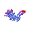 28089MC M: map data used to model this data C: citing same article ( |
|---|---|
| Similar structure data |
-Links
 PDBj
PDBj

- Assembly
| Deposited unit | 
|
|---|---|
| 1 |
|
-Components
| #1: Protein | Mass: 25550.348 Da / Num. of mol.: 5 / Mutation: S233Stop / Source method: isolated from a natural source / Source: (natural) Mus musculus (house mouse) / References: UniProt: P04925 |
|---|
-Experimental details
-Experiment
| Experiment | Method: ELECTRON MICROSCOPY |
|---|---|
| EM experiment | Aggregation state: FILAMENT / 3D reconstruction method: helical reconstruction |
- Sample preparation
| Component | Name: GPI anchorless underglycosylated 22L prion fibril / Type: COMPLEX / Entity ID: all / Source: NATURAL |
|---|---|
| Molecular weight | Experimental value: NO |
| Source (natural) | Organism: Mus musculus (house mouse) / Organ: Brain |
| Buffer solution | pH: 7.4 Details: Sample suspended in 20 mM Tris pH 7.4, 100 mM containing 0.02% amphipol 8-35 |
| Specimen | Embedding applied: NO / Shadowing applied: NO / Staining applied: NO / Vitrification applied: YES |
| Specimen support | Grid material: COPPER / Grid mesh size: 300 divisions/in. / Grid type: C-flat-1.2/1.3 |
| Vitrification | Instrument: LEICA EM GP / Cryogen name: ETHANE / Humidity: 90 % / Chamber temperature: 295 K |
- Electron microscopy imaging
| Experimental equipment |  Model: Titan Krios / Image courtesy: FEI Company |
|---|---|
| Microscopy | Model: FEI TITAN KRIOS |
| Electron gun | Electron source: FIELD EMISSION GUN / Accelerating voltage: 300 kV / Illumination mode: FLOOD BEAM |
| Electron lens | Mode: BRIGHT FIELDBright-field microscopy / Nominal magnification: 81000 X / Nominal defocus max: 3000 nm / Nominal defocus min: 500 nm |
| Specimen holder | Cryogen: NITROGEN / Specimen holder model: FEI TITAN KRIOS AUTOGRID HOLDER |
| Image recording | Electron dose: 57 e/Å2 / Film or detector model: GATAN K3 BIOQUANTUM (6k x 4k) / Num. of real images: 4449 |
| EM imaging optics | Energyfilter name: GIF Bioquantum / Energyfilter slit width: 20 eV |
- Processing
| EM software |
| ||||||||||||||||||||||||||||||||||||||||
|---|---|---|---|---|---|---|---|---|---|---|---|---|---|---|---|---|---|---|---|---|---|---|---|---|---|---|---|---|---|---|---|---|---|---|---|---|---|---|---|---|---|
| CTF correction | Type: PHASE FLIPPING AND AMPLITUDE CORRECTION | ||||||||||||||||||||||||||||||||||||||||
| Helical symmerty | Angular rotation/subunit: -0.565 ° / Axial rise/subunit: 4.75 Å / Axial symmetry: C1 | ||||||||||||||||||||||||||||||||||||||||
| Particle selection | Num. of particles selected: 226306 | ||||||||||||||||||||||||||||||||||||||||
| 3D reconstruction | Resolution: 3.2 Å / Resolution method: FSC 0.143 CUT-OFF / Num. of particles: 16409 Details: Pixel size was corrected in post processing from 1.1 A/pix to 1.045 A/pix to correct an error in the reported magnification pixel size. Symmetry type: HELICAL | ||||||||||||||||||||||||||||||||||||||||
| Atomic model building | Protocol: AB INITIO MODEL / Space: REAL |
