 Movie
Movie Controller


+ Open data

- Basic information
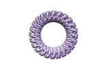
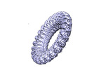
| Entry | Database: PDB / ID: 8efp | ||||||
|---|---|---|---|---|---|---|---|
| Title | CryoEM structure of GSDMB in complex with shigella IpaH7.8 | ||||||
 Components Components |
| ||||||
 Keywords Keywords | TRANSFERASE/LIPID BINDING PROTEIN / GSDMB /  GSDMD / pyroptosis / ubiquitination / ANTIMICROBIAL PROTEIN / TRANSFERASE-LIPID BINDING PROTEIN complex GSDMD / pyroptosis / ubiquitination / ANTIMICROBIAL PROTEIN / TRANSFERASE-LIPID BINDING PROTEIN complex | ||||||
| Function / homology |  Function and homology information Function and homology informationsymbiont-mediated suppression of host programmed cell death / cytotoxic T cell pyroptotic process / effector-mediated activation of programmed cell death in host / wide pore channel activity / killing by host of symbiont cells / cardiolipin binding / phosphatidylinositol-4-phosphate binding / phosphatidylserine binding / pyroptosis / phosphatidylinositol-4,5-bisphosphate binding ...symbiont-mediated suppression of host programmed cell death / cytotoxic T cell pyroptotic process / effector-mediated activation of programmed cell death in host / wide pore channel activity / killing by host of symbiont cells / cardiolipin binding / phosphatidylinositol-4-phosphate binding / phosphatidylserine binding / pyroptosis / phosphatidylinositol-4,5-bisphosphate binding / phospholipid binding / RING-type E3 ubiquitin transferase / ubiquitin protein ligase activity / ubiquitin-dependent protein catabolic process / killing of cells of another organism / defense response to Gram-negative bacterium / host cell cytoplasm / protein ubiquitination / defense response to bacterium / extracellular region / plasma membrane / cytoplasmSimilarity search - Function | ||||||
| Biological species |  Shigella flexneri (bacteria) Shigella flexneri (bacteria) Homo sapiens (human) Homo sapiens (human) | ||||||
| Method | ELECTRON MICROSCOPY / single particle reconstruction / cryo EM / Resolution: 3.8 Å | ||||||
 Authors Authors | Wang, C. / Ruan, J. | ||||||
| Funding support |  United States, 1items United States, 1items
| ||||||
 Citation Citation | Journal: Nature / Year: 2023 Title: Structural basis for GSDMB pore formation and its targeting by IpaH7.8. Authors: Chengliang Wang / Sonia Shivcharan / Tian Tian / Skylar Wright / Danyang Ma / JengYih Chang / Kunpeng Li / Kangkang Song / Chen Xu / Vijay A Rathinam / Jianbin Ruan / Abstract: Gasdermins (GSDMs) are pore-forming proteins that play critical roles in host defence through pyroptosis. Among GSDMs, GSDMB is unique owing to its distinct lipid-binding profile and a lack of ...Gasdermins (GSDMs) are pore-forming proteins that play critical roles in host defence through pyroptosis. Among GSDMs, GSDMB is unique owing to its distinct lipid-binding profile and a lack of consensus on its pyroptotic potential. Recently, GSDMB was shown to exhibit direct bactericidal activity through its pore-forming activity. Shigella, an intracellular, human-adapted enteropathogen, evades this GSDMB-mediated host defence by secreting IpaH7.8, a virulence effector that triggers ubiquitination-dependent proteasomal degradation of GSDMB. Here, we report the cryogenic electron microscopy structures of human GSDMB in complex with Shigella IpaH7.8 and the GSDMB pore. The structure of the GSDMB-IpaH7.8 complex identifies a motif of three negatively charged residues in GSDMB as the structural determinant recognized by IpaH7.8. Human, but not mouse, GSDMD contains this conserved motif, explaining the species specificity of IpaH7.8. The GSDMB pore structure shows the alternative splicing-regulated interdomain linker in GSDMB as a regulator of GSDMB pore formation. GSDMB isoforms with a canonical interdomain linker exhibit normal pyroptotic activity whereas other isoforms exhibit attenuated or no pyroptotic activity. Overall, this work sheds light on the molecular mechanisms of Shigella IpaH7.8 recognition and targeting of GSDMs and shows a structural determinant in GSDMB critical for its pyroptotic activity. | ||||||
| History |
|
- Structure visualization
| Structure viewer | Molecule: MolmilJmol/JSmol |
|---|
- Downloads & links
-Download
| PDBx/mmCIF format | 8efp.cif.gz | 120.6 KB | Display | PDBx/mmCIF format |
|---|---|---|---|---|
| PDB format | pdb8efp.ent.gz | 90.1 KB | Display | PDB format |
| PDBx/mmJSON format | 8efp.json.gz | Tree view | PDBx/mmJSON format | |
| Others |  Other downloads Other downloads |
-Validation report
| Arichive directory | https://data.pdbj.org/pub/pdb/validation_reports/ef/8efpftp://data.pdbj.org/pub/pdb/validation_reports/ef/8efp | HTTPS FTP |
|---|
-Related structure data
| Related structure data | 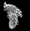 28087MC  8et1C  8et2C M: map data used to model this data C: citing same article ( |
|---|---|
| Similar structure data |
-Links
 PDBj
PDBj


- Assembly
| Deposited unit | 
|
|---|---|
| 1 |
|
-Components
| #1: Protein | Mass: 64602.949 Da / Num. of mol.: 1 Source method: isolated from a genetically manipulated source Source: (gene. exp.) Shigella flexneri (bacteria) / Gene: ipaH7.8, CP0078, pWR501_0084, SFLP133 / Production host: Escherichia coli (E. coli)References: UniProt: P18014, RING-type E3 ubiquitin transferase |
|---|---|
| #2: Protein | Mass: 47410.863 Da / Num. of mol.: 1 Source method: isolated from a genetically manipulated source Source: (gene. exp.) Homo sapiens (human) / Gene: GSDMB, GSDML, PP4052, PRO2521 / Production host: Escherichia coli (E. coli) / References: UniProt: Q8TAX9 |
-Experimental details
-Experiment
| Experiment | Method: ELECTRON MICROSCOPY |
|---|---|
| EM experiment | Aggregation state: PARTICLE / 3D reconstruction method: single particle reconstruction |
- Sample preparation
| Component |
| ||||||||||||||||||||||||
|---|---|---|---|---|---|---|---|---|---|---|---|---|---|---|---|---|---|---|---|---|---|---|---|---|---|
| Molecular weight | Experimental value: NO | ||||||||||||||||||||||||
| Source (natural) |
| ||||||||||||||||||||||||
| Source (recombinant) |
| ||||||||||||||||||||||||
| Buffer solution | pH: 7.5 | ||||||||||||||||||||||||
| Specimen | Embedding applied: NO / Shadowing applied: NO / Staining applied: NO / Vitrification applied: YES | ||||||||||||||||||||||||
| Specimen support | Grid material: COPPER / Grid mesh size: 400 divisions/in. / Grid type: Quantifoil R1.2/1.3 | ||||||||||||||||||||||||
| Vitrification | Cryogen name: ETHANE |
- Electron microscopy imaging
| Experimental equipment |  Model: Titan Krios / Image courtesy: FEI Company |
|---|---|
| Microscopy | Model: FEI TITAN KRIOS |
| Electron gun | Electron source: FIELD EMISSION GUN / Accelerating voltage: 300 kV / Illumination mode: SPOT SCAN |
| Electron lens | Mode: DIFFRACTION / Nominal defocus max: 2500 nm / Nominal defocus min: 800 nm |
| Image recording | Electron dose: 66.95 e/Å2 / Film or detector model: GATAN K3 BIOQUANTUM (6k x 4k) |
- Processing
| Software | Name: PHENIX / Version: 1.20.1_4487: / Classification: refinement | ||||||||||||||||||||||||
|---|---|---|---|---|---|---|---|---|---|---|---|---|---|---|---|---|---|---|---|---|---|---|---|---|---|
| CTF correction | Type: NONE | ||||||||||||||||||||||||
| 3D reconstruction | Resolution: 3.8 Å / Resolution method: FSC 0.143 CUT-OFF / Num. of particles: 113959 / Symmetry type: POINT | ||||||||||||||||||||||||
| Refine LS restraints |
|
