Journal: Nat Struct Mol Biol / Year: 2023 Title: Molecular mechanism of glutaminase activation through filamentation and the role of filaments in mitophagy protection. Authors: Douglas Adamoski / Marilia Meira Dias / Jose Edwin Neciosup Quesñay / Zhengyi Yang / Ievgeniia Zagoriy / Anna M Steyer / Camila Tanimoto Rodrigues / Alliny Cristiny da Silva Bastos / Bianca ...Authors: Douglas Adamoski / Marilia Meira Dias / Jose Edwin Neciosup Quesñay / Zhengyi Yang / Ievgeniia Zagoriy / Anna M Steyer / Camila Tanimoto Rodrigues / Alliny Cristiny da Silva Bastos / Bianca Novaes da Silva / Renna Karoline Eloi Costa / Flávia Mayumi Odahara de Abreu / Zeyaul Islam / Alexandre Cassago / Marin Gerard van Heel / Sílvio Roberto Consonni / Simone Mattei / Julia Mahamid / Rodrigo Villares Portugal / Andre Luis Berteli Ambrosio / Sandra Martha Gomes Dias / Abstract: Glutaminase (GLS), which deaminates glutamine to form glutamate, is a mitochondrial tetrameric protein complex. Although inorganic phosphate (Pi) is known to promote GLS filamentation and activation, ...Glutaminase (GLS), which deaminates glutamine to form glutamate, is a mitochondrial tetrameric protein complex. Although inorganic phosphate (Pi) is known to promote GLS filamentation and activation, the molecular basis of this mechanism is unknown. Here we aimed to determine the molecular mechanism of Pi-induced mouse GLS filamentation and its impact on mitochondrial physiology. Single-particle cryogenic electron microscopy revealed an allosteric mechanism in which Pi binding at the tetramer interface and the activation loop is coupled to direct nucleophile activation at the active site. The active conformation is prone to enzyme filamentation. Notably, human GLS filaments form inside tubulated mitochondria following glutamine withdrawal, as shown by in situ cryo-electron tomography of cells thinned by cryo-focused ion beam milling. Mitochondria with GLS filaments exhibit increased protection from mitophagy. We reveal roles of filamentous GLS in mitochondrial morphology and recycling.
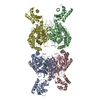
Conc.: 0.33 mg/ml / Embedding applied: NO / Shadowing applied: NO / Staining applied: NO / Vitrification applied: YES Details: In the presence of phosphate, GAC polymerizes and becomes polydisperse.
In the structure databanks used in Yorodumi, some data are registered as the other names, "COVID-19 virus" and "2019-nCoV". Here are the details of the virus and the list of structure data.
Jan 31, 2019. EMDB accession codes are about to change! (news from PDBe EMDB page)
EMDB accession codes are about to change! (news from PDBe EMDB page)
The allocation of 4 digits for EMDB accession codes will soon come to an end. Whilst these codes will remain in use, new EMDB accession codes will include an additional digit and will expand incrementally as the available range of codes is exhausted. The current 4-digit format prefixed with “EMD-” (i.e. EMD-XXXX) will advance to a 5-digit format (i.e. EMD-XXXXX), and so on. It is currently estimated that the 4-digit codes will be depleted around Spring 2019, at which point the 5-digit format will come into force.
The EM Navigator/Yorodumi systems omit the EMD- prefix.
Related info.:Q: What is EMD? / ID/Accession-code notation in Yorodumi/EM Navigator
Yorodumi is a browser for structure data from EMDB, PDB, SASBDB, etc.
This page is also the successor to EM Navigator detail page, and also detail information page/front-end page for Omokage search.
The word "yorodu" (or yorozu) is an old Japanese word meaning "ten thousand". "mi" (miru) is to see.
Related info.:EMDB / PDB / SASBDB / Comparison of 3 databanks / Yorodumi Search / Aug 31, 2016. New EM Navigator & Yorodumi / Yorodumi Papers / Jmol/JSmol / Function and homology information / Changes in new EM Navigator and Yorodumi
 Movie
Movie Controller


 Open data
 Basic information
 Components
 Keywords
 HYDROLASE /
  Function and homology information
 Authors
 Brazil, 1items
  Citation


 Structure visualization
 Downloads & links
 Other downloads
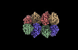
 PDBj
PDBj Assembly


 Mass: 94.971 Da / Num. of mol.: 8 / Source method: obtained synthetically / Formula: PO4 / Feature type: SUBJECT OF INVESTIGATION
 Sample preparation
 Electron microscopy imaging
 Processing