 Movie
Movie Controller

[English] 日本語
 Yorodumi
- PDB-8e5r: Schistosoma mansoni (Blood Fluke) Sulfotransferase/CIDD-0150610 C... -
+ Open data

- Basic information
| Entry | Database: PDB / ID: 8e5r | ||||||
|---|---|---|---|---|---|---|---|
| Title | Schistosoma mansoni (Blood Fluke) Sulfotransferase/CIDD-0150610 Complex | ||||||
 Components Components | Sulfotransferase oxamniquine resistance protein | ||||||
 Keywords Keywords |  TRANSFERASE / SULFOTRANSFERASE / PARASITE / DRUG RESISTANCE TRANSFERASE / SULFOTRANSFERASE / PARASITE / DRUG RESISTANCE | ||||||
| Function / homology | Sulfotransferase, S. mansonii-type / Sulfotransferase domain / transferase activity / P-loop containing nucleoside triphosphate hydrolase / ADENOSINE-3'-5'-DIPHOSPHATE / Chem-ULX / Sulfotransferase oxamniquine resistance protein Function and homology information Function and homology information | ||||||
| Biological species |  Schistosoma mansoni (invertebrata) Schistosoma mansoni (invertebrata) | ||||||
| Method | X-RAY DIFFRACTION / SYNCHROTRON / MOLECULAR REPLACEMENT / Resolution: 1.4 Å | ||||||
 Authors Authors | Taylor, A.B. / Alwan, S.N. | ||||||
| Funding support |  United States, 1items United States, 1items
| ||||||
 Citation Citation | Journal: Plos Pathog. / Year: 2023 Title: Oxamniquine derivatives overcome Praziquantel treatment limitations for Schistosomiasis. Authors: Alwan, S.N. / Taylor, A.B. / Rhodes, J. / Tidwell, M. / McHardy, S.F. / LoVerde, P.T. | ||||||
| History |
|
- Structure visualization
| Structure viewer | Molecule: MolmilJmol/JSmol |
|---|
- Downloads & links
-Download
| PDBx/mmCIF format | 8e5r.cif.gz | 139.6 KB | Display | PDBx/mmCIF format |
|---|---|---|---|---|
| PDB format | pdb8e5r.ent.gz | 98.3 KB | Display | PDB format |
| PDBx/mmJSON format | 8e5r.json.gz | Tree view | PDBx/mmJSON format | |
| Others |  Other downloads Other downloads |
-Validation report
| Arichive directory | https://data.pdbj.org/pub/pdb/validation_reports/e5/8e5rftp://data.pdbj.org/pub/pdb/validation_reports/e5/8e5r | HTTPS FTP |
|---|
-Related structure data
| Related structure data | 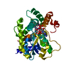 8e5qC 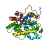 6bdrS S: Starting model for refinement C: citing same article ( |
|---|---|
| Similar structure data |
-Links
 PDBj
PDBj
- Assembly
| Deposited unit | 
| ||||||||||||
|---|---|---|---|---|---|---|---|---|---|---|---|---|---|
| 1 |
| ||||||||||||
| Unit cell |
| ||||||||||||
| Components on special symmetry positions |
|
-Components
| #1: Protein | Mass: 30080.547 Da / Num. of mol.: 1 Source method: isolated from a genetically manipulated source Source: (gene. exp.) Schistosoma mansoni (invertebrata) / Gene: SULT-OR, Smp_089320 / Plasmid: pAG8H / Production host:  Escherichia coli BL21(DE3) (bacteria) / Strain (production host): pLysS / References: UniProt: G4VLE5 Escherichia coli BL21(DE3) (bacteria) / Strain (production host): pLysS / References: UniProt: G4VLE5 |
|---|---|
| #2: Chemical | ChemComp-ULX / [  Mass: 540.577 Da / Num. of mol.: 1 / Source method: obtained synthetically / Formula: C29H31F3N4O3 / Feature type: SUBJECT OF INVESTIGATION Mass: 540.577 Da / Num. of mol.: 1 / Source method: obtained synthetically / Formula: C29H31F3N4O3 / Feature type: SUBJECT OF INVESTIGATION |
| #3: Chemical | ChemComp-A3P / Adenosine 3',5'-bisphosphate  Type: RNA linking / Mass: 427.201 Da / Num. of mol.: 1 / Source method: obtained synthetically / Formula: C10H15N5O10P2 Type: RNA linking / Mass: 427.201 Da / Num. of mol.: 1 / Source method: obtained synthetically / Formula: C10H15N5O10P2 |
| #4: Water | ChemComp-HOH / Water Mass: 18.015 Da / Num. of mol.: 222 / Source method: isolated from a natural source / Formula: H2O Mass: 18.015 Da / Num. of mol.: 222 / Source method: isolated from a natural source / Formula: H2O |
| Has ligand of interest | Y |
-Experimental details
-Experiment
| Experiment | Method: X-RAY DIFFRACTION / Number of used crystals: 1 |
|---|
- Sample preparation
| Crystal | Density Matthews: 2.51 Å3/Da / Density % sol: 50.98 % |
|---|---|
| Crystal grow | Temperature: 277 K / Method: vapor diffusion, sitting drop Details: MCSG-3 condition A1: 20% PEG 8000, 0.1 M HEPES:NaOH pH 7.5 |
-Data collection
| Diffraction | Mean temperature: 100 K / Serial crystal experiment: N |
|---|---|
| Diffraction source | Source: SYNCHROTRON / Site: APS / Beamline: 24-ID-E / Wavelength: 0.97918 Å |
| Detector | Type: DECTRIS EIGER X 16M / Detector: PIXEL / Date: Sep 23, 2020 |
| Radiation | Protocol: SINGLE WAVELENGTH / Monochromatic (M) / Laue (L): M / Scattering type: x-ray |
| Radiation wavelength | Wavelength: 0.97918 Å / Relative weight: 1 |
| Reflection | Resolution: 1.4→140.89 Å / Num. obs: 59038 / % possible obs: 97.6 % / Redundancy: 6 % / Biso Wilson estimate: 23.74 Å2 / CC1/2: 0.999 / Rpim(I) all: 0.022 / Net I/σ(I): 13.3 |
| Reflection shell | Resolution: 1.4→1.48 Å / Redundancy: 6.2 % / Mean I/σ(I) obs: 1 / Num. unique obs: 8718 / CC1/2: 0.515 / Rpim(I) all: 0.817 / % possible all: 99.9 |
- Processing
| Software |
| |||||||||||||||||||||||||||||||||||||||||||||||||||||||||||||||||||||||||||||||||||||||||||||||||||||||||
|---|---|---|---|---|---|---|---|---|---|---|---|---|---|---|---|---|---|---|---|---|---|---|---|---|---|---|---|---|---|---|---|---|---|---|---|---|---|---|---|---|---|---|---|---|---|---|---|---|---|---|---|---|---|---|---|---|---|---|---|---|---|---|---|---|---|---|---|---|---|---|---|---|---|---|---|---|---|---|---|---|---|---|---|---|---|---|---|---|---|---|---|---|---|---|---|---|---|---|---|---|---|---|---|---|---|---|
| Refinement | Method to determine structure: MOLECULAR REPLACEMENT Starting model: 6BDR Resolution: 1.4→53.84 Å / SU ML: 0.1726 / Cross valid method: FREE R-VALUE / σ(F): 1.34 / Phase error: 24.9043 Stereochemistry target values: GeoStd + Monomer Library + CDL v1.2
| |||||||||||||||||||||||||||||||||||||||||||||||||||||||||||||||||||||||||||||||||||||||||||||||||||||||||
| Solvent computation | Shrinkage radii: 0.9 Å / VDW probe radii: 1.1 Å / Solvent model: FLAT BULK SOLVENT MODEL | |||||||||||||||||||||||||||||||||||||||||||||||||||||||||||||||||||||||||||||||||||||||||||||||||||||||||
| Displacement parameters | Biso mean: 34.47 Å2 | |||||||||||||||||||||||||||||||||||||||||||||||||||||||||||||||||||||||||||||||||||||||||||||||||||||||||
| Refinement step | Cycle: LAST / Resolution: 1.4→53.84 Å
| |||||||||||||||||||||||||||||||||||||||||||||||||||||||||||||||||||||||||||||||||||||||||||||||||||||||||
| Refine LS restraints |
| |||||||||||||||||||||||||||||||||||||||||||||||||||||||||||||||||||||||||||||||||||||||||||||||||||||||||
| LS refinement shell |
|
