 Movie
Movie Controller

[English] 日本語
 Yorodumi
- PDB-8dkt: Crystal Structure of Septin1 - Septin2 heterocomplex from Drosoph... -
+ Open data

- Basic information
| Entry | Database: PDB / ID: 8dkt | ||||||
|---|---|---|---|---|---|---|---|
| Title | Crystal Structure of Septin1 - Septin2 heterocomplex from Drosophila melanogaster | ||||||
 Components Components |
| ||||||
 Keywords Keywords |  CELL CYCLE / Septin CELL CYCLE / Septin | ||||||
| Function / homology |  Function and homology information Function and homology informationgrowth of a germarium-derived egg chamber / imaginal disc development / cellularization / septin complex / imaginal disc-derived wing morphogenesis / cytoskeleton-dependent cytokinesis / septin ring / regulation of exocytosis / cell division site / cleavage furrow ...growth of a germarium-derived egg chamber / imaginal disc development / cellularization / septin complex / imaginal disc-derived wing morphogenesis / cytoskeleton-dependent cytokinesis / septin ring / regulation of exocytosis / cell division site / cleavage furrow / protein localization / spindle / microtubule cytoskeleton / synaptic vesicle / molecular adaptor activity / regulation of cell cycle / positive regulation of apoptotic process / GTPase activity / ubiquitin protein ligase binding / GTP binding / protein homodimerization activitySimilarity search - Function | ||||||
| Biological species |  Drosophila melanogaster (fruit fly) Drosophila melanogaster (fruit fly) | ||||||
| Method | X-RAY DIFFRACTION / SYNCHROTRON / MOLECULAR REPLACEMENT / molecular replacement / Resolution: 2.38 Å | ||||||
 Authors Authors | de Freitas, A.F. / Leonardo, D.A. / Cavini, I.A. / Pereira, H.M. / Garratt, R.C. | ||||||
| Funding support |  Brazil, 1items Brazil, 1items
| ||||||
 Citation Citation | Journal: Cytoskeleton (Hoboken) / Year: 2023 Title: Conservation and divergence of the G-interfaces of Drosophila melanogaster septins. Authors: de Freitas Fernandes, A. / Leonardo, D.A. / Cavini, I.A. / Rosa, H.V.D. / Vargas, J.A. / D'Muniz Pereira, H. / Nascimento, A.S. / Garratt, R.C. | ||||||
| History |
|
- Structure visualization
| Structure viewer | Molecule: MolmilJmol/JSmol |
|---|
- Downloads & links
-Download
| PDBx/mmCIF format | 8dkt.cif.gz | 230.2 KB | Display | PDBx/mmCIF format |
|---|---|---|---|---|
| PDB format | pdb8dkt.ent.gz | 181.5 KB | Display | PDB format |
| PDBx/mmJSON format | 8dkt.json.gz | Tree view | PDBx/mmJSON format | |
| Others |  Other downloads Other downloads |
-Validation report
| Arichive directory | https://data.pdbj.org/pub/pdb/validation_reports/dk/8dktftp://data.pdbj.org/pub/pdb/validation_reports/dk/8dkt | HTTPS FTP |
|---|
-Related structure data
| Related structure data | 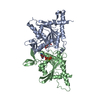 6upqS S: Starting model for refinement |
|---|---|
| Similar structure data |
-Links
 PDBj
PDBj- Assembly
| Deposited unit | 
| ||||||||
|---|---|---|---|---|---|---|---|---|---|
| 1 |
| ||||||||
| Unit cell |
|
-Components
-Protein , 2 types, 2 molecules AB
| #1: Protein | / DIFF6 protein homolog / Protein innocent bystander Mass: 31558.312 Da / Num. of mol.: 1 Source method: isolated from a genetically manipulated source Source: (gene. exp.) Drosophila melanogaster (fruit fly) / Gene: Sep1, Diff6, iby, CG1403 / Production host:  Escherichia coli (E. coli) / Strain (production host): DE3 / References: UniProt: P42207 Escherichia coli (E. coli) / Strain (production host): DE3 / References: UniProt: P42207 |
|---|---|
| #2: Protein | Mass: 32377.033 Da / Num. of mol.: 1 Source method: isolated from a genetically manipulated source Source: (gene. exp.) Drosophila melanogaster (fruit fly) / Gene: Sep2, CG4173 / Plasmid: pET Duet / Production host: Escherichia coli (E. coli) / Strain (production host): DE3 / References: UniProt: P54359 |
-Non-polymers , 4 types, 42 molecules 


| #3: Chemical | ChemComp-GDP / Guanosine diphosphate Type: RNA linking / Mass: 443.201 Da / Num. of mol.: 1 / Source method: obtained synthetically / Formula: C10H15N5O11P2 / Feature type: SUBJECT OF INVESTIGATION / Comment: GDP, energy-carrying molecule*YM Type: RNA linking / Mass: 443.201 Da / Num. of mol.: 1 / Source method: obtained synthetically / Formula: C10H15N5O11P2 / Feature type: SUBJECT OF INVESTIGATION / Comment: GDP, energy-carrying molecule*YM |
|---|---|
| #4: Chemical | ChemComp-GTP / Guanosine triphosphate Mass: 523.180 Da / Num. of mol.: 1 / Source method: obtained synthetically / Formula: C10H16N5O14P3 / Feature type: SUBJECT OF INVESTIGATION / Comment: GTP, energy-carrying molecule*YM Mass: 523.180 Da / Num. of mol.: 1 / Source method: obtained synthetically / Formula: C10H16N5O14P3 / Feature type: SUBJECT OF INVESTIGATION / Comment: GTP, energy-carrying molecule*YM |
| #5: Chemical | ChemComp-MG /  Mass: 24.305 Da / Num. of mol.: 1 / Source method: obtained synthetically / Formula: Mg / Feature type: SUBJECT OF INVESTIGATION Mass: 24.305 Da / Num. of mol.: 1 / Source method: obtained synthetically / Formula: Mg / Feature type: SUBJECT OF INVESTIGATION |
| #6: Water | ChemComp-HOH / WaterMass: 18.015 Da / Num. of mol.: 39 / Source method: isolated from a natural source / Formula: H2O |
-Details
| Has ligand of interest | Y |
|---|
-Experimental details
-Experiment
| Experiment | Method: X-RAY DIFFRACTION / Number of used crystals: 1 |
|---|
- Sample preparation
| Crystal | Density Matthews: 3.34 Å3/Da / Density % sol: 63.16 % |
|---|---|
| Crystal grow | Temperature: 291 K / Method: vapor diffusion, sitting drop / pH: 6.5 Details: 10% w/v PEG 8000, 20% v/v ethylene glycol; 0.1 M MES/imidazole pH 6.5; 0.02 M of each sodium formate, ammonium acetate, trisodium citrate, sodium potassium l-tartrate, sodium oxamate |
-Data collection
| Diffraction | Mean temperature: 100 K / Serial crystal experiment: N |
|---|---|
| Diffraction source | Source: SYNCHROTRON / Site: Diamond  / Beamline: I04 / Wavelength: 0.9795 Å / Beamline: I04 / Wavelength: 0.9795 Å |
| Detector | Type: DECTRIS EIGER2 XE 16M / Detector: PIXEL / Date: Feb 26, 2022 |
| Radiation | Protocol: SINGLE WAVELENGTH / Monochromatic (M) / Laue (L): M / Scattering type: x-ray |
| Radiation wavelength | Wavelength: 0.9795 Å / Relative weight: 1 |
| Reflection | Resolution: 2.38→42.56 Å / Num. obs: 15075 / % possible obs: 43.2 % / Redundancy: 13.4 % / Biso Wilson estimate: 57.29 Å2 / CC1/2: 1 / Rmerge(I) obs: 0.144 / Rpim(I) all: 0.041 / Rrim(I) all: 0.159 / Net I/σ(I): 11.1 |
| Reflection shell | Resolution: 2.38→2.747 Å / Redundancy: 12.3 % / Rmerge(I) obs: 1.548 / Mean I/σ(I) obs: 1.7 / Num. unique obs: 9267 / CC1/2: 0.632 / Rpim(I) all: 0.459 / Rrim(I) all: 1.616 / % possible all: 6.2 |
-Phasing
| Phasing | Method: molecular replacement |
|---|
- Processing
| Software |
| ||||||||||||||||||||||||||||||||||||||||||||||||||||||||||||||||||||||||||||||||||||||||||||||||||||||||||||||||||||||||||||||||||||||||||||||||||||||||||||||||||||||||||||||||||||||||||||||||||||||||||||||||||||||||||||||||||||||||||||||||||||||||||||||||||||||||||||||||||||||||||||||||||||||||||||
|---|---|---|---|---|---|---|---|---|---|---|---|---|---|---|---|---|---|---|---|---|---|---|---|---|---|---|---|---|---|---|---|---|---|---|---|---|---|---|---|---|---|---|---|---|---|---|---|---|---|---|---|---|---|---|---|---|---|---|---|---|---|---|---|---|---|---|---|---|---|---|---|---|---|---|---|---|---|---|---|---|---|---|---|---|---|---|---|---|---|---|---|---|---|---|---|---|---|---|---|---|---|---|---|---|---|---|---|---|---|---|---|---|---|---|---|---|---|---|---|---|---|---|---|---|---|---|---|---|---|---|---|---|---|---|---|---|---|---|---|---|---|---|---|---|---|---|---|---|---|---|---|---|---|---|---|---|---|---|---|---|---|---|---|---|---|---|---|---|---|---|---|---|---|---|---|---|---|---|---|---|---|---|---|---|---|---|---|---|---|---|---|---|---|---|---|---|---|---|---|---|---|---|---|---|---|---|---|---|---|---|---|---|---|---|---|---|---|---|---|---|---|---|---|---|---|---|---|---|---|---|---|---|---|---|---|---|---|---|---|---|---|---|---|---|---|---|---|---|---|---|---|---|---|---|---|---|---|---|---|---|---|---|---|---|---|---|---|---|---|---|---|---|---|---|---|---|---|---|---|---|---|---|---|---|---|---|---|---|---|---|---|---|---|---|---|---|---|---|---|---|---|
| Refinement | Method to determine structure: MOLECULAR REPLACEMENT Starting model: 6UPQ Resolution: 2.38→42.56 Å / SU ML: 0.29 / Cross valid method: THROUGHOUT / σ(F): 1.35 / Phase error: 32.19 / Stereochemistry target values: ML
| ||||||||||||||||||||||||||||||||||||||||||||||||||||||||||||||||||||||||||||||||||||||||||||||||||||||||||||||||||||||||||||||||||||||||||||||||||||||||||||||||||||||||||||||||||||||||||||||||||||||||||||||||||||||||||||||||||||||||||||||||||||||||||||||||||||||||||||||||||||||||||||||||||||||||||||
| Solvent computation | Shrinkage radii: 0.9 Å / VDW probe radii: 1.11 Å / Solvent model: FLAT BULK SOLVENT MODEL | ||||||||||||||||||||||||||||||||||||||||||||||||||||||||||||||||||||||||||||||||||||||||||||||||||||||||||||||||||||||||||||||||||||||||||||||||||||||||||||||||||||||||||||||||||||||||||||||||||||||||||||||||||||||||||||||||||||||||||||||||||||||||||||||||||||||||||||||||||||||||||||||||||||||||||||
| Displacement parameters | Biso max: 172.89 Å2 / Biso mean: 69.4938 Å2 / Biso min: 22.82 Å2 | ||||||||||||||||||||||||||||||||||||||||||||||||||||||||||||||||||||||||||||||||||||||||||||||||||||||||||||||||||||||||||||||||||||||||||||||||||||||||||||||||||||||||||||||||||||||||||||||||||||||||||||||||||||||||||||||||||||||||||||||||||||||||||||||||||||||||||||||||||||||||||||||||||||||||||||
| Refinement step | Cycle: final / Resolution: 2.38→42.56 Å
| ||||||||||||||||||||||||||||||||||||||||||||||||||||||||||||||||||||||||||||||||||||||||||||||||||||||||||||||||||||||||||||||||||||||||||||||||||||||||||||||||||||||||||||||||||||||||||||||||||||||||||||||||||||||||||||||||||||||||||||||||||||||||||||||||||||||||||||||||||||||||||||||||||||||||||||
| LS refinement shell | Refine-ID: X-RAY DIFFRACTION / Rfactor Rfree error: 0 / Total num. of bins used: 5
| ||||||||||||||||||||||||||||||||||||||||||||||||||||||||||||||||||||||||||||||||||||||||||||||||||||||||||||||||||||||||||||||||||||||||||||||||||||||||||||||||||||||||||||||||||||||||||||||||||||||||||||||||||||||||||||||||||||||||||||||||||||||||||||||||||||||||||||||||||||||||||||||||||||||||||||
| Refinement TLS params. | Method: refined / Refine-ID: X-RAY DIFFRACTION
| ||||||||||||||||||||||||||||||||||||||||||||||||||||||||||||||||||||||||||||||||||||||||||||||||||||||||||||||||||||||||||||||||||||||||||||||||||||||||||||||||||||||||||||||||||||||||||||||||||||||||||||||||||||||||||||||||||||||||||||||||||||||||||||||||||||||||||||||||||||||||||||||||||||||||||||
| Refinement TLS group |
|
