 Movie
Movie Controller

[English] 日本語
 Yorodumi
- PDB-8de5: Structure of glyceraldehyde-3-phosphate dehydrogenase from Paraco... -
+ Open data

- Basic information
| Entry | Database: PDB / ID: 8de5 | ||||||
|---|---|---|---|---|---|---|---|
| Title | Structure of glyceraldehyde-3-phosphate dehydrogenase from Paracoccidioides lutzii | ||||||
 Components Components | Glyceraldehyde-3-phosphate dehydrogenase Glyceraldehyde 3-phosphate dehydrogenase Glyceraldehyde 3-phosphate dehydrogenase | ||||||
 Keywords Keywords | OXIDOREDUCTASE / Glyceraldehyde-3-Phosphate Dehydrogenase / Paracoccidioides lutzii / Paracoccidioidomycosis / Aldonic Sugar Acid | ||||||
| Function / homology |  Function and homology informationglyceraldehyde-3-phosphate dehydrogenase (phosphorylating) / glyceraldehyde-3-phosphate dehydrogenase (NAD+) (phosphorylating) activity / glycolytic process / glucose metabolic process / NAD binding / NADP binding / cytoplasm Function and homology informationglyceraldehyde-3-phosphate dehydrogenase (phosphorylating) / glyceraldehyde-3-phosphate dehydrogenase (NAD+) (phosphorylating) activity / glycolytic process / glucose metabolic process / NAD binding / NADP binding / cytoplasmSimilarity search - Function | ||||||
| Biological species |  Paracoccidioides lutzii Pb01 (fungus) Paracoccidioides lutzii Pb01 (fungus) | ||||||
| Method | X-RAY DIFFRACTION / SYNCHROTRON / MOLECULAR REPLACEMENT / Resolution: 2.02 Å | ||||||
 Authors Authors | Hernandez-Prieto, J.H. / Martini, V.P. / Iulek, J. | ||||||
| Funding support |  Brazil, 1items Brazil, 1items
| ||||||
 Citation Citation | Journal: Biochimie / Year: 2023 Title: Structure of glyceraldehyde-3-phosphate dehydrogenase from Paracoccidioides lutzii in complex with an aldonic sugar acid. Authors: Hernandez-Prieto, J.H. / Martini, V.P. / Iulek, J. | ||||||
| History |
|
- Structure visualization
| Structure viewer | Molecule: MolmilJmol/JSmol |
|---|
- Downloads & links
-Download
| PDBx/mmCIF format | 8de5.cif.gz | 192.1 KB | Display | PDBx/mmCIF format |
|---|---|---|---|---|
| PDB format | pdb8de5.ent.gz | 126.1 KB | Display | PDB format |
| PDBx/mmJSON format | 8de5.json.gz | Tree view | PDBx/mmJSON format | |
| Others |  Other downloads Other downloads |
-Validation report
| Arichive directory | https://data.pdbj.org/pub/pdb/validation_reports/de/8de5ftp://data.pdbj.org/pub/pdb/validation_reports/de/8de5 | HTTPS FTP |
|---|
-Related structure data
| Related structure data | 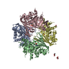 7u4sS S: Starting model for refinement |
|---|---|
| Similar structure data |
-Links
 PDBj
PDBj
- Assembly
| Deposited unit | 
| ||||||||||||
|---|---|---|---|---|---|---|---|---|---|---|---|---|---|
| 1 | 
| ||||||||||||
| Unit cell |
| ||||||||||||
| Components on special symmetry positions |
|
-Components
-Protein , 1 types, 1 molecules A
| #1: Protein | Glyceraldehyde 3-phosphate dehydrogenase / GAPDH Mass: 36548.363 Da / Num. of mol.: 1 Source method: isolated from a genetically manipulated source Source: (gene. exp.) Paracoccidioides lutzii Pb01 (fungus) / Strain: ATCC MYA-826 / Pb01 / Gene: GPD, PAAG_08468 / Plasmid: pET-28a(+) / Production host:  Escherichia coli (E. coli) Escherichia coli (E. coli)References: UniProt: Q8X1X3, glyceraldehyde-3-phosphate dehydrogenase (phosphorylating) |
|---|
-Non-polymers , 5 types, 423 molecules 


| #2: Chemical | ChemComp-NAD / Nicotinamide adenine dinucleotide Mass: 663.425 Da / Num. of mol.: 1 / Source method: obtained synthetically / Formula: C21H27N7O14P2 / Feature type: SUBJECT OF INVESTIGATION / Comment: NAD*YM Mass: 663.425 Da / Num. of mol.: 1 / Source method: obtained synthetically / Formula: C21H27N7O14P2 / Feature type: SUBJECT OF INVESTIGATION / Comment: NAD*YM | ||
|---|---|---|---|
| #3: Chemical | ChemComp-J0M /  Mass: 196.155 Da / Num. of mol.: 1 / Source method: obtained synthetically / Formula: C6H12O7 / Feature type: SUBJECT OF INVESTIGATION Mass: 196.155 Da / Num. of mol.: 1 / Source method: obtained synthetically / Formula: C6H12O7 / Feature type: SUBJECT OF INVESTIGATION | ||
| #4: Chemical | ChemComp-SO4 / Sulfate Mass: 96.063 Da / Num. of mol.: 1 / Source method: obtained synthetically / Formula: SO4 / Feature type: SUBJECT OF INVESTIGATION Mass: 96.063 Da / Num. of mol.: 1 / Source method: obtained synthetically / Formula: SO4 / Feature type: SUBJECT OF INVESTIGATION | ||
| #5: Chemical | ChemComp-GOL / Glycerol Mass: 92.094 Da / Num. of mol.: 6 / Source method: obtained synthetically / Formula: C3H8O3 / Feature type: SUBJECT OF INVESTIGATION Mass: 92.094 Da / Num. of mol.: 6 / Source method: obtained synthetically / Formula: C3H8O3 / Feature type: SUBJECT OF INVESTIGATION#6: Water | ChemComp-HOH / | WaterMass: 18.015 Da / Num. of mol.: 414 / Source method: isolated from a natural source / Formula: H2O |
-Details
| Has ligand of interest | Y |
|---|
-Experimental details
-Experiment
| Experiment | Method: X-RAY DIFFRACTION / Number of used crystals: 1 |
|---|
- Sample preparation
| Crystal | Density Matthews: 3.67 Å3/Da / Density % sol: 66.5 % / Description: Tetragonal trapezohedral |
|---|---|
| Crystal grow | Temperature: 291.15 K / Method: vapor diffusion, sitting drop / pH: 9.3 / Details: 0.2 M ammonium sulfate, 25.5% w/v PEG4000 |
-Data collection
| Diffraction | Mean temperature: 100 K / Serial crystal experiment: N |
|---|---|
| Diffraction source | Source: SYNCHROTRON / Site: LNLS SIRUS / Beamline: MANACA / Wavelength: 0.97718 Å |
| Detector | Type: DECTRIS PILATUS 2M / Detector: PIXEL / Date: Nov 2, 2021 |
| Radiation | Protocol: SINGLE WAVELENGTH / Monochromatic (M) / Laue (L): M / Scattering type: x-ray |
| Radiation wavelength | Wavelength: 0.97718 Å / Relative weight: 1 |
| Reflection | Resolution: 2.02→83.94 Å / Num. obs: 37329 / % possible obs: 100 % / Redundancy: 56.6 % / Biso Wilson estimate: 35.362 Å2 / CC1/2: 0.998 / Rmerge(I) obs: 0.606 / Rpim(I) all: 0.074 / Rrim(I) all: 0.611 / Net I/σ(I): 10.4 |
| Reflection shell | Resolution: 2.02→2.05 Å / Redundancy: 26.6 % / Rmerge(I) obs: 3.767 / Mean I/σ(I) obs: 1.1 / Num. unique obs: 1814 / CC1/2: 0.372 / Rpim(I) all: 0.742 / Rrim(I) all: 3.84 / % possible all: 100 |
- Processing
| Software |
| |||||||||||||||||||||||||||||||||||||||||||||||||||||||||||||||||||||||||||||||||||||||||||||||||||||||||
|---|---|---|---|---|---|---|---|---|---|---|---|---|---|---|---|---|---|---|---|---|---|---|---|---|---|---|---|---|---|---|---|---|---|---|---|---|---|---|---|---|---|---|---|---|---|---|---|---|---|---|---|---|---|---|---|---|---|---|---|---|---|---|---|---|---|---|---|---|---|---|---|---|---|---|---|---|---|---|---|---|---|---|---|---|---|---|---|---|---|---|---|---|---|---|---|---|---|---|---|---|---|---|---|---|---|---|
| Refinement | Method to determine structure: MOLECULAR REPLACEMENT Starting model: PDB entry 7U4S Resolution: 2.02→83.94 Å / SU ML: 0.2587 / Cross valid method: FREE R-VALUE / σ(F): 1.35 / Phase error: 22.0854 Stereochemistry target values: GeoStd + Monomer Library + CDL v1.2
| |||||||||||||||||||||||||||||||||||||||||||||||||||||||||||||||||||||||||||||||||||||||||||||||||||||||||
| Solvent computation | Shrinkage radii: 1.2 Å / VDW probe radii: 1.3 Å / Solvent model: FLAT BULK SOLVENT MODEL | |||||||||||||||||||||||||||||||||||||||||||||||||||||||||||||||||||||||||||||||||||||||||||||||||||||||||
| Displacement parameters | Biso mean: 49.41 Å2 | |||||||||||||||||||||||||||||||||||||||||||||||||||||||||||||||||||||||||||||||||||||||||||||||||||||||||
| Refinement step | Cycle: LAST / Resolution: 2.02→83.94 Å
| |||||||||||||||||||||||||||||||||||||||||||||||||||||||||||||||||||||||||||||||||||||||||||||||||||||||||
| Refine LS restraints |
| |||||||||||||||||||||||||||||||||||||||||||||||||||||||||||||||||||||||||||||||||||||||||||||||||||||||||
| LS refinement shell |
| |||||||||||||||||||||||||||||||||||||||||||||||||||||||||||||||||||||||||||||||||||||||||||||||||||||||||
| Refinement TLS params. | Method: refined / Origin x: 0.677084236769 Å / Origin y: -39.9064479659 Å / Origin z: -24.9261160368 Å
| |||||||||||||||||||||||||||||||||||||||||||||||||||||||||||||||||||||||||||||||||||||||||||||||||||||||||
| Refinement TLS group | Selection details: chain A |
