 Movie
Movie Controller

[English] 日本語
 Yorodumi
- PDB-7u4s: Structure of Glyceraldehyde-3-Phosphate Dehydrogenase from Candid... -
+ Open data

- Basic information
| Entry | Database: PDB / ID: 7u4s | ||||||
|---|---|---|---|---|---|---|---|
| Title | Structure of Glyceraldehyde-3-Phosphate Dehydrogenase from Candida albicans | ||||||
 Components Components | Glyceraldehyde-3-phosphate dehydrogenase Glyceraldehyde 3-phosphate dehydrogenase Glyceraldehyde 3-phosphate dehydrogenase | ||||||
 Keywords Keywords | OXIDOREDUCTASE / Glyceraldehyde-3-Phosphate Dehydrogenase | ||||||
| Function / homology |  Function and homology informationglyceraldehyde-3-phosphate dehydrogenase (phosphorylating) / glyceraldehyde-3-phosphate dehydrogenase (NAD+) (phosphorylating) activity / glycolytic process / cell wall organization / glucose metabolic process / NAD binding / NADP binding / extracellular region / cytoplasm Function and homology informationglyceraldehyde-3-phosphate dehydrogenase (phosphorylating) / glyceraldehyde-3-phosphate dehydrogenase (NAD+) (phosphorylating) activity / glycolytic process / cell wall organization / glucose metabolic process / NAD binding / NADP binding / extracellular region / cytoplasmSimilarity search - Function | ||||||
| Biological species |  Candida albicans WO-1 (yeast) Candida albicans WO-1 (yeast) | ||||||
| Method | X-RAY DIFFRACTION / SYNCHROTRON / MOLECULAR REPLACEMENT / Resolution: 2.68 Å | ||||||
 Authors Authors | Miranda, R.R. / Silva, M. / Iulek, J. | ||||||
| Funding support |  Brazil, 1items Brazil, 1items
| ||||||
 Citation Citation | Journal: Chem. Data Coll. / Year: 2022 Title: Expression, purification, crystallization and structure of Glyceraldehyde-3-Phosphate Dehydrogenase from Candida albicans, main causative agent of candidiasis Authors: Miranda, R.R. / Silva, M. / Iulek, J. | ||||||
| History |
|
- Structure visualization
| Structure viewer | Molecule: MolmilJmol/JSmol |
|---|
- Downloads & links
-Download
| PDBx/mmCIF format | 7u4s.cif.gz | 592.1 KB | Display | PDBx/mmCIF format |
|---|---|---|---|---|
| PDB format | pdb7u4s.ent.gz | 408.6 KB | Display | PDB format |
| PDBx/mmJSON format | 7u4s.json.gz | Tree view | PDBx/mmJSON format | |
| Others |  Other downloads Other downloads |
-Validation report
| Arichive directory | https://data.pdbj.org/pub/pdb/validation_reports/u4/7u4sftp://data.pdbj.org/pub/pdb/validation_reports/u4/7u4s | HTTPS FTP |
|---|
-Related structure data
| Related structure data | 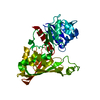 4iq8S S: Starting model for refinement |
|---|---|
| Similar structure data |
-Links
 PDBj
PDBj
- Assembly
| Deposited unit | 
| ||||||||||||
|---|---|---|---|---|---|---|---|---|---|---|---|---|---|
| 1 |
| ||||||||||||
| Unit cell |
|
-Components
| #1: Protein | Glyceraldehyde 3-phosphate dehydrogenase / GAPDH Mass: 35924.852 Da / Num. of mol.: 4 Source method: isolated from a genetically manipulated source Source: (gene. exp.) Candida albicans WO-1 (yeast) / Strain: WO-1 / Gene: TDH1, GAP, TDH3, CAWG_02997 / Production host:  Escherichia coli (E. coli) Escherichia coli (E. coli)References: UniProt: Q92211, glyceraldehyde-3-phosphate dehydrogenase (phosphorylating)#2: Chemical | Glycerol  Mass: 92.094 Da / Num. of mol.: 3 / Source method: obtained synthetically / Formula: C3H8O3 Mass: 92.094 Da / Num. of mol.: 3 / Source method: obtained synthetically / Formula: C3H8O3#3: Water | ChemComp-HOH / | Water Mass: 18.015 Da / Num. of mol.: 462 / Source method: isolated from a natural source / Formula: H2O Mass: 18.015 Da / Num. of mol.: 462 / Source method: isolated from a natural source / Formula: H2OHas ligand of interest | N | |
|---|
-Experimental details
-Experiment
| Experiment | Method: X-RAY DIFFRACTION / Number of used crystals: 1 |
|---|
- Sample preparation
| Crystal | Density Matthews: 2.78 Å3/Da / Density % sol: 55.76 % |
|---|---|
| Crystal grow | Temperature: 291.15 K / Method: vapor diffusion, hanging drop Details: 20% w/v PEG6000 0.1 M citrate, pH 4.0, 1.0 M lithium chloride |
-Data collection
| Diffraction | Mean temperature: 100 K / Serial crystal experiment: N |
|---|---|
| Diffraction source | Source: SYNCHROTRON / Site: SOLEIL  / Beamline: PROXIMA 1 / Wavelength: 0.97856 Å / Beamline: PROXIMA 1 / Wavelength: 0.97856 Å |
| Detector | Type: DECTRIS EIGER X 16M / Detector: PIXEL / Date: Sep 9, 2020 |
| Radiation | Protocol: SINGLE WAVELENGTH / Monochromatic (M) / Laue (L): M / Scattering type: x-ray |
| Radiation wavelength | Wavelength: 0.97856 Å / Relative weight: 1 |
| Reflection | Resolution: 2.68→72.52 Å / Num. obs: 44083 / % possible obs: 100 % / Redundancy: 13 % / Biso Wilson estimate: 40.72 Å2 / CC1/2: 0.966 / Rpim(I) all: 0.183 / Net I/σ(I): 4.7 |
| Reflection shell | Resolution: 2.68→2.75 Å / Redundancy: 7 % / Num. unique obs: 3258 / CC1/2: 0.4 / Rpim(I) all: 2.205 / % possible all: 100 |
- Processing
| Software |
| |||||||||||||||||||||||||||||||||||||||||||||||||||||||||||||||||||||||||||||||||||||||||||||||||||||||||||||||||||||||||||||
|---|---|---|---|---|---|---|---|---|---|---|---|---|---|---|---|---|---|---|---|---|---|---|---|---|---|---|---|---|---|---|---|---|---|---|---|---|---|---|---|---|---|---|---|---|---|---|---|---|---|---|---|---|---|---|---|---|---|---|---|---|---|---|---|---|---|---|---|---|---|---|---|---|---|---|---|---|---|---|---|---|---|---|---|---|---|---|---|---|---|---|---|---|---|---|---|---|---|---|---|---|---|---|---|---|---|---|---|---|---|---|---|---|---|---|---|---|---|---|---|---|---|---|---|---|---|---|
| Refinement | Method to determine structure: MOLECULAR REPLACEMENT Starting model: PDB entry 4IQ8 Resolution: 2.68→72.52 Å / SU ML: 0.4565 / Cross valid method: FREE R-VALUE / σ(F): 1.34 / Phase error: 32.4152 Stereochemistry target values: GeoStd + Monomer Library + CDL v1.2
| |||||||||||||||||||||||||||||||||||||||||||||||||||||||||||||||||||||||||||||||||||||||||||||||||||||||||||||||||||||||||||||
| Solvent computation | Shrinkage radii: 1.1 Å / VDW probe radii: 1.2 Å / Solvent model: FLAT BULK SOLVENT MODEL | |||||||||||||||||||||||||||||||||||||||||||||||||||||||||||||||||||||||||||||||||||||||||||||||||||||||||||||||||||||||||||||
| Displacement parameters | Biso mean: 53.29 Å2 | |||||||||||||||||||||||||||||||||||||||||||||||||||||||||||||||||||||||||||||||||||||||||||||||||||||||||||||||||||||||||||||
| Refinement step | Cycle: LAST / Resolution: 2.68→72.52 Å
| |||||||||||||||||||||||||||||||||||||||||||||||||||||||||||||||||||||||||||||||||||||||||||||||||||||||||||||||||||||||||||||
| Refine LS restraints |
| |||||||||||||||||||||||||||||||||||||||||||||||||||||||||||||||||||||||||||||||||||||||||||||||||||||||||||||||||||||||||||||
| LS refinement shell |
| |||||||||||||||||||||||||||||||||||||||||||||||||||||||||||||||||||||||||||||||||||||||||||||||||||||||||||||||||||||||||||||
| Refinement TLS params. | Method: refined / Refine-ID: X-RAY DIFFRACTION
| |||||||||||||||||||||||||||||||||||||||||||||||||||||||||||||||||||||||||||||||||||||||||||||||||||||||||||||||||||||||||||||
| Refinement TLS group | Refine-ID: X-RAY DIFFRACTION / Auth seq-ID: 1 - 335
|
