 Movie
Movie Controller

[English] 日本語
 Yorodumi
- PDB-8dcq: CRYSTAL STRUCTURE OF HIV-1 LM/HT CLADE A/E CRF01 GP120 CORE IN CO... -
+ Open data

- Basic information
| Entry | Database: PDB / ID: 8dcq | ||||||
|---|---|---|---|---|---|---|---|
| Title | CRYSTAL STRUCTURE OF HIV-1 LM/HT CLADE A/E CRF01 GP120 CORE IN COMPLEX WITH YIR-821 | ||||||
 Components Components | HIV-1 LM/HT Clade A/E CRF01 gp120 core | ||||||
 Keywords Keywords |  VIRAL PROTEIN / HIV-1 GP120 / CLADE A/E CF01 / VIRAL PROTEIN-INHIBITOR COMPLEX VIRAL PROTEIN / HIV-1 GP120 / CLADE A/E CF01 / VIRAL PROTEIN-INHIBITOR COMPLEX | ||||||
| Function / homology | Gp120 core superfamily / Envelope glycoprotein GP120 / Human immunodeficiency virus 1, envelope glycoprotein Gp120 / viral envelope / Chem-R5K / clade A/E 93TH057 HIV-1 gp120 core Function and homology information Function and homology information | ||||||
| Biological species |   Human immunodeficiency virus 1 Human immunodeficiency virus 1 | ||||||
| Method | X-RAY DIFFRACTION / SYNCHROTRON / MOLECULAR REPLACEMENT / Resolution: 3.2 Å | ||||||
 Authors Authors | Tolbert, W.D. / Pazgier, M. | ||||||
| Funding support |  United States, 1items United States, 1items
| ||||||
 Citation Citation | Journal: J.Virol. / Year: 2023 Title: Characterization of a Novel CD4 Mimetic Compound YIR-821 against HIV-1 Clinical Isolates. Authors: Matsumoto, K. / Kuwata, T. / Tolbert, W.D. / Richard, J. / Ding, S. / Prevost, J. / Takahama, S. / Judicate, G.P. / Ueno, T. / Nakata, H. / Kobayakawa, T. / Tsuji, K. / Tamamura, H. / Smith ...Authors: Matsumoto, K. / Kuwata, T. / Tolbert, W.D. / Richard, J. / Ding, S. / Prevost, J. / Takahama, S. / Judicate, G.P. / Ueno, T. / Nakata, H. / Kobayakawa, T. / Tsuji, K. / Tamamura, H. / Smith 3rd, A.B. / Pazgier, M. / Finzi, A. / Matsushita, S. | ||||||
| History |
|
- Structure visualization
| Structure viewer | Molecule: MolmilJmol/JSmol |
|---|
- Downloads & links
-Download
| PDBx/mmCIF format | 8dcq.cif.gz | 90.1 KB | Display | PDBx/mmCIF format |
|---|---|---|---|---|
| PDB format | pdb8dcq.ent.gz | 65.2 KB | Display | PDB format |
| PDBx/mmJSON format | 8dcq.json.gz | Tree view | PDBx/mmJSON format | |
| Others |  Other downloads Other downloads |
-Validation report
| Arichive directory | https://data.pdbj.org/pub/pdb/validation_reports/dc/8dcqftp://data.pdbj.org/pub/pdb/validation_reports/dc/8dcq | HTTPS FTP |
|---|
-Related structure data
| Related structure data | 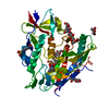 6onfS S: Starting model for refinement |
|---|---|
| Similar structure data |
-Links
 PDBj
PDBj


- Assembly
| Deposited unit | 
| ||||||||
|---|---|---|---|---|---|---|---|---|---|
| 1 |
| ||||||||
| Unit cell |
|
-Components
| #1: Protein | Mass: 39466.750 Da / Num. of mol.: 1 / Mutation: H61Y Q105H V108I H375T N474D I475M K476R Source method: isolated from a genetically manipulated source Source: (gene. exp.) Human immunodeficiency virus 1 / Gene: HIV-1 Env / Cell (production host): HEK 293 GNT1- / Production host:  Homo sapiens (human) / References: UniProt: A0A0M3KKW9 Homo sapiens (human) / References: UniProt: A0A0M3KKW9 | ||||||
|---|---|---|---|---|---|---|---|
| #2: Sugar | ChemComp-NAG / N-Acetylglucosamine  Type: D-saccharide, beta linking / Mass: 221.208 Da / Num. of mol.: 10 Type: D-saccharide, beta linking / Mass: 221.208 Da / Num. of mol.: 10Source method: isolated from a genetically manipulated source Formula: C8H15NO6 #3: Chemical | ChemComp-R5K / |   Mass: 534.094 Da / Num. of mol.: 1 / Source method: obtained synthetically / Formula: C26H40ClN7O3 / Feature type: SUBJECT OF INVESTIGATION Mass: 534.094 Da / Num. of mol.: 1 / Source method: obtained synthetically / Formula: C26H40ClN7O3 / Feature type: SUBJECT OF INVESTIGATION#4: Chemical | ChemComp-EPE / | HEPES  Mass: 238.305 Da / Num. of mol.: 1 / Source method: obtained synthetically / Formula: C8H18N2O4S / Comment: pH buffer*YM Mass: 238.305 Da / Num. of mol.: 1 / Source method: obtained synthetically / Formula: C8H18N2O4S / Comment: pH buffer*YMHas ligand of interest | Y | |
-Experimental details
-Experiment
| Experiment | Method: X-RAY DIFFRACTION / Number of used crystals: 1 |
|---|
- Sample preparation
| Crystal | Density Matthews: 2.45 Å3/Da / Density % sol: 49.7 % |
|---|---|
| Crystal grow | Temperature: 294 K / Method: vapor diffusion, hanging drop / pH: 7.5 / Details: 10% PEG 1500 5% PEG 400 0.1 M HEPES pH 7.5 |
-Data collection
| Diffraction | Mean temperature: 100 K / Serial crystal experiment: N |
|---|---|
| Diffraction source | Source: SYNCHROTRON / Site: SSRL / Beamline: BL12-2 / Wavelength: 0.97946 Å |
| Detector | Type: DECTRIS PILATUS 6M / Detector: PIXEL / Date: Nov 9, 2018 |
| Radiation | Monochromator: Si(111) / Protocol: SINGLE WAVELENGTH / Monochromatic (M) / Laue (L): M / Scattering type: x-ray |
| Radiation wavelength | Wavelength: 0.97946 Å / Relative weight: 1 |
| Reflection | Resolution: 3.2→50 Å / Num. obs: 8078 / % possible obs: 72.3 % / Redundancy: 3.3 % / CC1/2: 0.94 / Rpim(I) all: 0.169 / Rsym value: 0.276 / Net I/σ(I): 2.1 |
| Reflection shell | Resolution: 3.2→3.37 Å / Mean I/σ(I) obs: 0.5 / Num. unique obs: 722 / CC1/2: 0.464 / Rpim(I) all: 0.745 / % possible all: 75.3 |
- Processing
| Software |
| ||||||||||||||||||||||||||||
|---|---|---|---|---|---|---|---|---|---|---|---|---|---|---|---|---|---|---|---|---|---|---|---|---|---|---|---|---|---|
| Refinement | Method to determine structure: MOLECULAR REPLACEMENT Starting model: 6ONF Resolution: 3.2→43.69 Å / SU ML: 0.38 / Cross valid method: FREE R-VALUE / σ(F): 0.08 / Phase error: 24.62 / Stereochemistry target values: ML
| ||||||||||||||||||||||||||||
| Solvent computation | Shrinkage radii: 0.9 Å / VDW probe radii: 1.11 Å / Solvent model: FLAT BULK SOLVENT MODEL | ||||||||||||||||||||||||||||
| Refinement step | Cycle: LAST / Resolution: 3.2→43.69 Å
| ||||||||||||||||||||||||||||
| Refine LS restraints |
| ||||||||||||||||||||||||||||
| LS refinement shell |
|
