 Movie
Movie Controller

[English] 日本語
 Yorodumi
- PDB-8d1w: Crystal structure of Plasmodium falciparum GRP78 in complex with ... -
+ Open data

- Basic information
| Entry | Database: PDB / ID: 8d1w | ||||||
|---|---|---|---|---|---|---|---|
| Title | Crystal structure of Plasmodium falciparum GRP78 in complex with (2R,3R,4S,5R)-2-(6-amino-8-((2-chlorobenzyl)amino)-9H-purin-9-yl)-5-(hydroxymethyl)tetrahydrofuran-3,4-diol | ||||||
 Components Components | Chaperone DnaK | ||||||
 Keywords Keywords |  CHAPERONE / ATPase CHAPERONE / ATPase | ||||||
| Function / homology |  Function and homology information Function and homology informationATP-dependent protein folding chaperone / endoplasmic reticulum / ATP bindingSimilarity search - Function | ||||||
| Biological species |  Plasmodium falciparum (malaria parasite P. falciparum) Plasmodium falciparum (malaria parasite P. falciparum) | ||||||
| Method | X-RAY DIFFRACTION / SYNCHROTRON / MOLECULAR REPLACEMENT / Resolution: 2.1 Å | ||||||
 Authors Authors | Mrozek, A. / Chen, Y. / Antoshchenko, T. / Park, H.W. / Smil, D. / Zepeda, C.A.V. | ||||||
| Funding support |  United States, 1items United States, 1items
| ||||||
 Citation Citation | Journal: Frontiers in Molecular Bioscience / Year: 2022 Title: Targeting Plasmodium falciparum GRP78: nucleoside analogues as agents against the malaria chaperone Authors: Mrozek, A. / Park, H.W. | ||||||
| History |
|
- Structure visualization
| Structure viewer | Molecule: MolmilJmol/JSmol |
|---|
- Downloads & links
-Download
| PDBx/mmCIF format | 8d1w.cif.gz | 97.4 KB | Display | PDBx/mmCIF format |
|---|---|---|---|---|
| PDB format | pdb8d1w.ent.gz | 70.8 KB | Display | PDB format |
| PDBx/mmJSON format | 8d1w.json.gz | Tree view | PDBx/mmJSON format | |
| Others |  Other downloads Other downloads |
-Validation report
| Arichive directory | https://data.pdbj.org/pub/pdb/validation_reports/d1/8d1wftp://data.pdbj.org/pub/pdb/validation_reports/d1/8d1w | HTTPS FTP |
|---|
-Related structure data
| Related structure data | 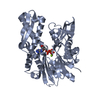 5umbS S: Starting model for refinement |
|---|---|
| Similar structure data |
-Links
 PDBj
PDBj
- Assembly
| Deposited unit | 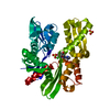
| ||||||||
|---|---|---|---|---|---|---|---|---|---|
| 1 |
| ||||||||
| Unit cell |
|
-Components
| #1: Protein | Mass: 44360.379 Da / Num. of mol.: 1 Source method: isolated from a genetically manipulated source Source: (gene. exp.) Plasmodium falciparum (malaria parasite P. falciparum)Strain: isolate Palo Alto / Uganda / Gene: PFUGPA_02944 / Production host:  Escherichia coli (E. coli) / References: UniProt: W4J0F1 Escherichia coli (E. coli) / References: UniProt: W4J0F1 | ||||
|---|---|---|---|---|---|
| #2: Chemical | ChemComp-HFY /   Mass: 406.824 Da / Num. of mol.: 1 / Source method: obtained synthetically / Formula: C17H19ClN6O4 / Feature type: SUBJECT OF INVESTIGATION Mass: 406.824 Da / Num. of mol.: 1 / Source method: obtained synthetically / Formula: C17H19ClN6O4 / Feature type: SUBJECT OF INVESTIGATION | ||||
| #3: Chemical | Sulfate  Mass: 96.063 Da / Num. of mol.: 2 / Source method: obtained synthetically / Formula: SO4 Mass: 96.063 Da / Num. of mol.: 2 / Source method: obtained synthetically / Formula: SO4#4: Water | ChemComp-HOH / | Water Mass: 18.015 Da / Num. of mol.: 266 / Source method: isolated from a natural source / Formula: H2O Mass: 18.015 Da / Num. of mol.: 266 / Source method: isolated from a natural source / Formula: H2OHas ligand of interest | Y | |
-Experimental details
-Experiment
| Experiment | Method: X-RAY DIFFRACTION / Number of used crystals: 1 |
|---|
- Sample preparation
| Crystal | Density Matthews: 3.59 Å3/Da / Density % sol: 65.69 % |
|---|---|
| Crystal grow | Temperature: 298 K / Method: vapor diffusion, sitting drop / pH: 5.6 Details: 0.7 M (NH4)2SO4, 1.2 M Li2SO4, 0.1 M NaCitrate, pH 5.6 |
-Data collection
| Diffraction | Mean temperature: 100 K / Serial crystal experiment: N |
|---|---|
| Diffraction source | Source: SYNCHROTRON / Site: APS / Beamline: 19-ID / Wavelength: 0.9792 Å |
| Detector | Type: DECTRIS PILATUS3 6M / Detector: PIXEL / Date: Mar 6, 2018 |
| Radiation | Protocol: SINGLE WAVELENGTH / Monochromatic (M) / Laue (L): M / Scattering type: x-ray |
| Radiation wavelength | Wavelength: 0.9792 Å / Relative weight: 1 |
| Reflection | Resolution: 2.1→50 Å / Num. obs: 36996 / % possible obs: 100 % / Redundancy: 18.9 % / Rrim(I) all: 0.1 / Rsym value: 0.09 / Net I/σ(I): 28.9 |
| Reflection shell | Resolution: 2.1→2.23 Å / Num. unique obs: 5825 / Rrim(I) all: 0.51 / Rsym value: 0.49 / % possible all: 100 |
- Processing
| Software |
| ||||||||||||||||||||||||||||||||||||||||||||||||||||||||||||||||||||||||||||||||||||||||||||||||||||||||||||||||||||||||||||||||||||||||||||||||||||||||||||||||||||||||||||||||||||||
|---|---|---|---|---|---|---|---|---|---|---|---|---|---|---|---|---|---|---|---|---|---|---|---|---|---|---|---|---|---|---|---|---|---|---|---|---|---|---|---|---|---|---|---|---|---|---|---|---|---|---|---|---|---|---|---|---|---|---|---|---|---|---|---|---|---|---|---|---|---|---|---|---|---|---|---|---|---|---|---|---|---|---|---|---|---|---|---|---|---|---|---|---|---|---|---|---|---|---|---|---|---|---|---|---|---|---|---|---|---|---|---|---|---|---|---|---|---|---|---|---|---|---|---|---|---|---|---|---|---|---|---|---|---|---|---|---|---|---|---|---|---|---|---|---|---|---|---|---|---|---|---|---|---|---|---|---|---|---|---|---|---|---|---|---|---|---|---|---|---|---|---|---|---|---|---|---|---|---|---|---|---|---|---|
| Refinement | Method to determine structure: MOLECULAR REPLACEMENT Starting model: 5UMB Resolution: 2.1→45.66 Å / Cor.coef. Fo:Fc: 0.95 / Cor.coef. Fo:Fc free: 0.939 / SU B: 3.481 / SU ML: 0.093 / Cross valid method: THROUGHOUT / ESU R: 0.156 / ESU R Free: 0.144 / Stereochemistry target values: MAXIMUM LIKELIHOOD / Details: HYDROGENS HAVE BEEN ADDED IN THE RIDING POSITIONS
| ||||||||||||||||||||||||||||||||||||||||||||||||||||||||||||||||||||||||||||||||||||||||||||||||||||||||||||||||||||||||||||||||||||||||||||||||||||||||||||||||||||||||||||||||||||||
| Solvent computation | Ion probe radii: 0.8 Å / Shrinkage radii: 0.8 Å / VDW probe radii: 1.2 Å / Solvent model: MASK | ||||||||||||||||||||||||||||||||||||||||||||||||||||||||||||||||||||||||||||||||||||||||||||||||||||||||||||||||||||||||||||||||||||||||||||||||||||||||||||||||||||||||||||||||||||||
| Displacement parameters | Biso mean: 33.561 Å2
| ||||||||||||||||||||||||||||||||||||||||||||||||||||||||||||||||||||||||||||||||||||||||||||||||||||||||||||||||||||||||||||||||||||||||||||||||||||||||||||||||||||||||||||||||||||||
| Refinement step | Cycle: 1 / Resolution: 2.1→45.66 Å
| ||||||||||||||||||||||||||||||||||||||||||||||||||||||||||||||||||||||||||||||||||||||||||||||||||||||||||||||||||||||||||||||||||||||||||||||||||||||||||||||||||||||||||||||||||||||
| Refine LS restraints |
|