 Movie
Movie Controller

[English] 日本語
 Yorodumi
- PDB-8cz9: Crystal Structure of the E372K LNK SH2 Domain mutant in Complex w... -
+ Open data

- Basic information
| Entry | Database: PDB / ID: 8cz9 | ||||||
|---|---|---|---|---|---|---|---|
| Title | Crystal Structure of the E372K LNK SH2 Domain mutant in Complex with a JAK2 pY813 Phosphopeptide | ||||||
 Components Components |
| ||||||
 Keywords Keywords |  SIGNALING PROTEIN / LNK / SH2B3 / JAK/STAT / MPNs SIGNALING PROTEIN / LNK / SH2B3 / JAK/STAT / MPNs | ||||||
| Function / homology |  Function and homology information Function and homology informationnegative regulation of Kit signaling pathway / monocyte homeostasis / thrombopoietin-mediated signaling pathway / stem cell factor receptor binding / negative regulation of sprouting angiogenesis / negative regulation of chemokine-mediated signaling pathway / negative regulation of response to cytokine stimulus / regulation of regulatory T cell differentiation / negative regulation of tyrosine phosphorylation of STAT protein / negative regulation of receptor signaling pathway via JAK-STAT ...negative regulation of Kit signaling pathway / monocyte homeostasis / thrombopoietin-mediated signaling pathway / stem cell factor receptor binding / negative regulation of sprouting angiogenesis / negative regulation of chemokine-mediated signaling pathway / negative regulation of response to cytokine stimulus / regulation of regulatory T cell differentiation / negative regulation of tyrosine phosphorylation of STAT protein / negative regulation of receptor signaling pathway via JAK-STAT / neutrophil homeostasis / negative regulation of platelet aggregation / cellular response to chemokine / cellular response to interleukin-3 / embryonic hemopoiesis / megakaryocyte development / erythrocyte development / phosphate ion binding / hemopoiesis / negative regulation of phosphatidylinositol 3-kinase/protein kinase B signal transduction / hematopoietic stem cell differentiation / protein tyrosine kinase binding / negative regulation of MAP kinase activity / signaling receptor complex adaptor activity / cell differentiation / intracellular signal transduction / negative regulation of cell population proliferation / protein-containing complex bindingSimilarity search - Function | ||||||
| Biological species |  Mus musculus (house mouse) Mus musculus (house mouse) Homo sapiens (human) Homo sapiens (human) | ||||||
| Method | X-RAY DIFFRACTION / SYNCHROTRON / MOLECULAR REPLACEMENT / Resolution: 1.65 Å | ||||||
 Authors Authors | Morris, R. / Kershaw, N.J. / Babon, J.J. | ||||||
| Funding support |  Australia, 1items Australia, 1items
| ||||||
 Citation Citation | Journal: J.Exp.Med. / Year: 2024 Title: Rare SH2B3 coding variants in lupus patients impair B cell tolerance and predispose to autoimmunity. Authors: Zhang, Y. / Morris, R. / Brown, G.J. / Lorenzo, A.M.D. / Meng, X. / Kershaw, N.J. / Kiridena, P. / Burgio, G. / Gross, S. / Cappello, J.Y. / Shen, Q. / Wang, H. / Turnbull, C. / Lea-Henry, T. ...Authors: Zhang, Y. / Morris, R. / Brown, G.J. / Lorenzo, A.M.D. / Meng, X. / Kershaw, N.J. / Kiridena, P. / Burgio, G. / Gross, S. / Cappello, J.Y. / Shen, Q. / Wang, H. / Turnbull, C. / Lea-Henry, T. / Stanley, M. / Yu, Z. / Ballard, F.D. / Chuah, A. / Lee, J.C. / Hatch, A.M. / Enders, A. / Masters, S.L. / Headley, A.P. / Trnka, P. / Mallon, D. / Fletcher, J.T. / Walters, G.D. / Sestan, M. / Jelusic, M. / Cook, M.C. / Athanasopoulos, V. / Fulcher, D.A. / Babon, J.J. / Vinuesa, C.G. / Ellyard, J.I. | ||||||
| History |
|
- Structure visualization
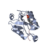
| Structure viewer | Molecule: MolmilJmol/JSmol |
|---|
- Downloads & links
-Download
| PDBx/mmCIF format | 8cz9.cif.gz | 41.2 KB | Display | PDBx/mmCIF format |
|---|---|---|---|---|
| PDB format | pdb8cz9.ent.gz | 25.6 KB | Display | PDB format |
| PDBx/mmJSON format | 8cz9.json.gz | Tree view | PDBx/mmJSON format | |
| Others |  Other downloads Other downloads |
-Validation report
| Arichive directory | https://data.pdbj.org/pub/pdb/validation_reports/cz/8cz9ftp://data.pdbj.org/pub/pdb/validation_reports/cz/8cz9 | HTTPS FTP |
|---|
-Related structure data
| Related structure data | 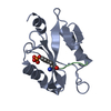 7r8wS S: Starting model for refinement |
|---|---|
| Similar structure data |
-Links
 PDBj
PDBj- Assembly
| Deposited unit | 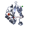
| |||||||||
|---|---|---|---|---|---|---|---|---|---|---|
| 1 |
| |||||||||
| Unit cell |
| |||||||||
| Components on special symmetry positions |
|
-Components
| #1: Protein | Mass: 12581.616 Da / Num. of mol.: 1 / Mutation: E372K Source method: isolated from a genetically manipulated source Source: (gene. exp.) Mus musculus (house mouse) / Gene: Sh2b3, Lnk / Production host:  Escherichia coli (E. coli) / References: UniProt: P50745 Escherichia coli (E. coli) / References: UniProt: P50745 |
|---|---|
| #2: Protein/peptide | Mass: 1059.018 Da / Num. of mol.: 1 / Source method: obtained synthetically / Source: (synth.) Homo sapiens (human) |
| #3: Chemical | ChemComp-CL / Chloride  Mass: 35.453 Da / Num. of mol.: 1 / Source method: obtained synthetically / Formula: Cl / Feature type: SUBJECT OF INVESTIGATION Mass: 35.453 Da / Num. of mol.: 1 / Source method: obtained synthetically / Formula: Cl / Feature type: SUBJECT OF INVESTIGATION |
| #4: Water | ChemComp-HOH / Water Mass: 18.015 Da / Num. of mol.: 76 / Source method: isolated from a natural source / Formula: H2O Mass: 18.015 Da / Num. of mol.: 76 / Source method: isolated from a natural source / Formula: H2O |
| Has ligand of interest | Y |
-Experimental details
-Experiment
| Experiment | Method: X-RAY DIFFRACTION / Number of used crystals: 1 |
|---|
- Sample preparation
| Crystal | Density Matthews: 2.5 Å3/Da / Density % sol: 50.79 % |
|---|---|
| Crystal grow | Temperature: 281.15 K / Method: vapor diffusion, hanging drop / Details: 18% PEG 8000, 0.05 M MgAc , 0.1 M Tris ph 8.5 |
-Data collection
| Diffraction | Mean temperature: 100 K / Serial crystal experiment: N |
|---|---|
| Diffraction source | Source: SYNCHROTRON / Site: Australian Synchrotron / Beamline: MX2 / Wavelength: 0.95373 Å |
| Detector | Type: DECTRIS EIGER X 16M / Detector: PIXEL / Date: Feb 23, 2018 |
| Radiation | Protocol: SINGLE WAVELENGTH / Monochromatic (M) / Laue (L): M / Scattering type: x-ray |
| Radiation wavelength | Wavelength: 0.95373 Å / Relative weight: 1 |
| Reflection | Resolution: 1.64→35.9 Å / Num. obs: 111266 / % possible obs: 99 % / Redundancy: 6.8 % / CC1/2: 0.99 / Net I/σ(I): 20.45 |
| Reflection shell | Resolution: 1.65→1.7 Å / Mean I/σ(I) obs: 2.49 / Num. unique obs: 11000 / CC1/2: 0.818 / Rrim(I) all: 0.76 |
- Processing
| Software |
| |||||||||||||||||||||||||||||||||||||||||||||||||||||||||||||||||||||||||||||||||||||||||||
|---|---|---|---|---|---|---|---|---|---|---|---|---|---|---|---|---|---|---|---|---|---|---|---|---|---|---|---|---|---|---|---|---|---|---|---|---|---|---|---|---|---|---|---|---|---|---|---|---|---|---|---|---|---|---|---|---|---|---|---|---|---|---|---|---|---|---|---|---|---|---|---|---|---|---|---|---|---|---|---|---|---|---|---|---|---|---|---|---|---|---|---|---|
| Refinement | Method to determine structure: MOLECULAR REPLACEMENT Starting model: 7R8W Resolution: 1.65→35.9 Å / SU ML: 0.21 / Cross valid method: THROUGHOUT / σ(F): 1.38 / Phase error: 21.63 / Stereochemistry target values: ML
| |||||||||||||||||||||||||||||||||||||||||||||||||||||||||||||||||||||||||||||||||||||||||||
| Solvent computation | Shrinkage radii: 0.9 Å / VDW probe radii: 1.11 Å / Solvent model: FLAT BULK SOLVENT MODEL | |||||||||||||||||||||||||||||||||||||||||||||||||||||||||||||||||||||||||||||||||||||||||||
| Displacement parameters | Biso max: 70.04 Å2 / Biso mean: 32.7311 Å2 / Biso min: 17.26 Å2 | |||||||||||||||||||||||||||||||||||||||||||||||||||||||||||||||||||||||||||||||||||||||||||
| Refinement step | Cycle: final / Resolution: 1.65→35.9 Å
| |||||||||||||||||||||||||||||||||||||||||||||||||||||||||||||||||||||||||||||||||||||||||||
| LS refinement shell | Refine-ID: X-RAY DIFFRACTION / Rfactor Rfree error: 0 / Total num. of bins used: 12
|
