 Movie
Movie Controller

[English] 日本語
 Yorodumi
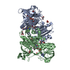
- PDB-8ct4: Cryo-EM structure of Mtb Lpd bound to inhibitor complex with 2-((... -
+ Open data

- Basic information
| Entry | Database: PDB / ID: 8ct4 | ||||||||||||
|---|---|---|---|---|---|---|---|---|---|---|---|---|---|
| Title | Cryo-EM structure of Mtb Lpd bound to inhibitor complex with 2-((2-cyano-N,5-dimethyl-1H-indole)-7-sulfonamido)-N-(4-(oxetan-3-yl)-3,4-dihydro-2H-benzo[b] [1,4]oxazin-7-yl)acetamide | ||||||||||||
 Components Components | Dihydrolipoyl dehydrogenase Dihydrolipoamide dehydrogenase Dihydrolipoamide dehydrogenase | ||||||||||||
 Keywords Keywords | OXIDOREDUCTASE/INHIBITOR / flavoprotein / glycolysis / redox-active center / OXIDOREDUCTASE-INHIBITOR complex | ||||||||||||
| Function / homology |  Function and homology information Function and homology informationCell redox homeostasis / dihydrolipoyl dehydrogenase / dihydrolipoyl dehydrogenase activity / pyruvate dehydrogenase complex / NADH binding / disulfide oxidoreductase activity / zymogen binding / oxidoreductase activity, acting on NAD(P)H, quinone or similar compound as acceptor / antioxidant activity / Prevention of phagosomal-lysosomal fusion ...Cell redox homeostasis / dihydrolipoyl dehydrogenase / dihydrolipoyl dehydrogenase activity / pyruvate dehydrogenase complex / NADH binding / disulfide oxidoreductase activity / zymogen binding / oxidoreductase activity, acting on NAD(P)H, quinone or similar compound as acceptor / antioxidant activity / Prevention of phagosomal-lysosomal fusion / tricarboxylic acid cycle / cell redox homeostasis / glycolytic process / flavin adenine dinucleotide binding / extracellular region / plasma membrane / cytosolSimilarity search - Function | ||||||||||||
| Biological species |   Mycobacterium tuberculosis (bacteria) Mycobacterium tuberculosis (bacteria) | ||||||||||||
| Method | ELECTRON MICROSCOPY / single particle reconstruction / cryo EM / Resolution: 2.17 Å | ||||||||||||
 Authors Authors | Kochanczyk, T. / Arango, N. / Lima, C.D. | ||||||||||||
| Funding support |  United States, 3items United States, 3items
| ||||||||||||
 Citation Citation | Journal: Not published Title: Cryo-EM structure of Mtb Lpd bound to the inhibitor 2-((2-cyano-N,5-dimethyl-1H-indole)-7-sulfonamido)-N-(4-(oxetan-3-yl)-3,4-dihydro-2H-benzo[b] [1,4]oxazin-7-yl)acetamide at 2.17 Angstrom resolution Authors: Kochanczyk, T. / Arango, N. / Michino, M. / Sun, S. / Ginn, J. / Bryk, R. / Nathan, C. / Lima, C.D. | ||||||||||||
| History |
|
- Structure visualization
| Structure viewer | Molecule: MolmilJmol/JSmol |
|---|
- Downloads & links
-Download
| PDBx/mmCIF format | 8ct4.cif.gz | 358.2 KB | Display | PDBx/mmCIF format |
|---|---|---|---|---|
| PDB format | pdb8ct4.ent.gz | 300.2 KB | Display | PDB format |
| PDBx/mmJSON format | 8ct4.json.gz | Tree view | PDBx/mmJSON format | |
| Others |  Other downloads Other downloads |
-Validation report
| Arichive directory | https://data.pdbj.org/pub/pdb/validation_reports/ct/8ct4ftp://data.pdbj.org/pub/pdb/validation_reports/ct/8ct4 | HTTPS FTP |
|---|
-Related structure data
| Related structure data |  26981MC M: map data used to model this data C: citing same article ( |
|---|---|
| Similar structure data |
-Links
 PDBj
PDBj


- Assembly
| Deposited unit | 
|
|---|---|
| 1 |
|
-Components
| #1: Protein | Dihydrolipoamide dehydrogenase / LPD / Component of peroxynitrite reductase/peroxidase complex / Component of PNR/P / ...LPD / Component of peroxynitrite reductase/peroxidase complex / Component of PNR/P / Dihydrolipoamide dehydrogenase / E3 component of alpha-ketoacid dehydrogenase complexes Mass: 49437.035 Da / Num. of mol.: 2 Source method: isolated from a genetically manipulated source Source: (gene. exp.) Mycobacterium tuberculosis (bacteria) / Strain: ATCC 25618 / H37Rv / Gene: lpdC, lpd, Rv0462, MTV038.06 / Production host: Escherichia coli BL21(DE3) (bacteria) / References: UniProt: P9WHH9, dihydrolipoyl dehydrogenase#2: Chemical | Flavin adenine dinucleotide  Mass: 785.550 Da / Num. of mol.: 2 / Source method: obtained synthetically / Formula: C27H33N9O15P2 / Comment: FAD*YM Mass: 785.550 Da / Num. of mol.: 2 / Source method: obtained synthetically / Formula: C27H33N9O15P2 / Comment: FAD*YM#3: Chemical | 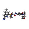  Mass: 495.551 Da / Num. of mol.: 2 / Source method: obtained synthetically / Formula: C24H25N5O5S / Feature type: SUBJECT OF INVESTIGATION Mass: 495.551 Da / Num. of mol.: 2 / Source method: obtained synthetically / Formula: C24H25N5O5S / Feature type: SUBJECT OF INVESTIGATION#4: Water | ChemComp-HOH / | Water Mass: 18.015 Da / Num. of mol.: 665 / Source method: isolated from a natural source / Formula: H2O Mass: 18.015 Da / Num. of mol.: 665 / Source method: isolated from a natural source / Formula: H2OHas ligand of interest | Y | |
|---|
-Experimental details
-Experiment
| Experiment | Method: ELECTRON MICROSCOPY |
|---|---|
| EM experiment | Aggregation state: PARTICLE / 3D reconstruction method: single particle reconstruction |
- Sample preparation
| Component | Name: Dihydrolipoyl dehydrogenase in complex with 2-((2-cyano-N,5-dimethyl-1H-indole)-7-sulfonamido)-N-(4-(oxetan-3-yl)-3,4-dihydro-2H-benzo[b] [1,4]oxazin-7-yl)acetamideDihydrolipoamide dehydrogenase Type: COMPLEX Details: 2-((2-cyano-N,5-dimethyl-1H-indole)-7-sulfonamido)-N-(4-(oxetan-3-yl)-3,4-dihydro-2H-benzo[b] [1,4]oxazin-7-yl)acetamide provided by the Tri-Institutional Therapeutics Discovery Institute ...Details: 2-((2-cyano-N,5-dimethyl-1H-indole)-7-sulfonamido)-N-(4-(oxetan-3-yl)-3,4-dihydro-2H-benzo[b] [1,4]oxazin-7-yl)acetamide provided by the Tri-Institutional Therapeutics Discovery Institute (tritdi.org) contact: John Ginn (jginn@tritdi.org) Entity ID: #1 / Source: RECOMBINANT |
|---|---|
| Molecular weight | Value: 0.099 MDa / Experimental value: NO |
| Source (natural) | Organism: Mycobacterium tuberculosis (bacteria) / Strain: ATCC 25618 / H37Rv |
| Source (recombinant) | Organism: Escherichia coli BL21(DE3) (bacteria) |
| Buffer solution | pH: 8 / Details: 20 mM Tris-HCl, pH 8.0, 87.5 mM NaCl, 0.05% IGEPAL |
| Specimen | Conc.: 3 mg/ml / Embedding applied: NO / Shadowing applied: NO / Staining applied: NO / Vitrification applied: YES |
| Specimen support | Grid material: GOLD / Grid mesh size: 300 divisions/in. / Grid type: UltrAuFoil R1.2/1.3 |
| Vitrification | Instrument: FEI VITROBOT MARK IV / Cryogen name: ETHANE Details: Temperature 22 C, humidity 100%, Wait time 8s, Blot time: 3.5s |
- Electron microscopy imaging
| Experimental equipment |  Model: Titan Krios / Image courtesy: FEI Company |
|---|---|
| Microscopy | Model: FEI TITAN KRIOS |
| Electron gun | Electron source: FIELD EMISSION GUN / Accelerating voltage: 300 kV / Illumination mode: FLOOD BEAM |
| Electron lens | Mode: BRIGHT FIELDBright-field microscopy / Nominal magnification: 22500 X / Nominal defocus max: 3000 nm / Nominal defocus min: 1000 nm / Cs: 2.7 mm |
| Specimen holder | Cryogen: NITROGEN / Specimen holder model: FEI TITAN KRIOS AUTOGRID HOLDER |
| Image recording | Average exposure time: 4 sec. / Electron dose: 70.66 e/Å2 / Detector mode: SUPER-RESOLUTION / Film or detector model: GATAN K3 (6k x 4k) / Num. of grids imaged: 1 / Num. of real images: 10748 |
- Processing
| EM software |
| ||||||||||||||||||||||||||||||||
|---|---|---|---|---|---|---|---|---|---|---|---|---|---|---|---|---|---|---|---|---|---|---|---|---|---|---|---|---|---|---|---|---|---|
| CTF correction | Type: PHASE FLIPPING AND AMPLITUDE CORRECTION | ||||||||||||||||||||||||||||||||
| Symmetry | Point symmetry: C2 (2 fold cyclic) | ||||||||||||||||||||||||||||||||
| 3D reconstruction | Resolution: 2.17 Å / Resolution method: FSC 0.143 CUT-OFF / Num. of particles: 1177958 / Symmetry type: POINT | ||||||||||||||||||||||||||||||||
| Atomic model building | Protocol: FLEXIBLE FIT / Space: REAL / Target criteria: Correlation coefficient | ||||||||||||||||||||||||||||||||
| Atomic model building | PDB-ID: 7KMY Pdb chain-ID: AB |
