 Movie
Movie Controller

[English] 日本語
 Yorodumi
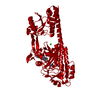
- PDB-8co5: The surface-engineered photosensory module (PAS-GAF-PHY) of the b... -
+ Open data

- Basic information
| Entry | Database: PDB / ID: 8co5 | ||||||
|---|---|---|---|---|---|---|---|
| Title | The surface-engineered photosensory module (PAS-GAF-PHY) of the bacterial phytochrome Agp1 (AtBphP1) in the Pr form with parallel dimer formation | ||||||
 Components Components | Bacteriophytochrome protein | ||||||
 Keywords Keywords |  SIGNALING PROTEIN / BILIN CHROMOPHORE / PARALLEL DIMER / PHYTOCHROME / PHOTORECEPTOR SIGNALING PROTEIN / BILIN CHROMOPHORE / PARALLEL DIMER / PHYTOCHROME / PHOTORECEPTOR | ||||||
| Function / homology |  Function and homology information Function and homology informationred or far-red light photoreceptor activity / red, far-red light phototransduction / osmosensory signaling via phosphorelay pathway / protein histidine kinase activity / detection of visible light / phosphorelay response regulator activity / protein kinase activator activity / histidine kinase / phosphorelay sensor kinase activity / regulation of DNA-templated transcription / metal ion bindingSimilarity search - Function | ||||||
| Biological species |  Agrobacterium fabrum str. C58 (bacteria) Agrobacterium fabrum str. C58 (bacteria) | ||||||
| Method | X-RAY DIFFRACTION / SYNCHROTRON / MOLECULAR REPLACEMENT / Resolution: 2.42 Å | ||||||
 Authors Authors | Schmidt, A. / Sauthof, L. / Krauss, N. / Scheerer, P. | ||||||
| Funding support |  Germany, 1items Germany, 1items
| ||||||
 Citation Citation | Journal: To Be Published Title: Crystal structures of a bacterial phytochrome exhibiting a group-subgroup relationship reveal pronounced flexibility of the photosensory core module in the Pr state Authors: Scheerer, P. / Schmidt, A. / Nagano, S. / Qureshi, B.M. / Szczepek, M. / Sauthof, L. / Michael, N. / Inomata, K. / Lamparter, T. / Krauss, N. | ||||||
| History |
|
- Structure visualization
| Structure viewer | Molecule: MolmilJmol/JSmol |
|---|
- Downloads & links
-Download
| PDBx/mmCIF format | 8co5.cif.gz | 203 KB | Display | PDBx/mmCIF format |
|---|---|---|---|---|
| PDB format | pdb8co5.ent.gz | 160.2 KB | Display | PDB format |
| PDBx/mmJSON format | 8co5.json.gz | Tree view | PDBx/mmJSON format | |
| Others |  Other downloads Other downloads |
-Validation report
| Arichive directory | https://data.pdbj.org/pub/pdb/validation_reports/co/8co5ftp://data.pdbj.org/pub/pdb/validation_reports/co/8co5 | HTTPS FTP |
|---|
-Related structure data
| Related structure data | |
|---|---|
| Similar structure data |


-Links
 PDBj
PDBj


- Assembly
| Deposited unit | 
| |||||||||
|---|---|---|---|---|---|---|---|---|---|---|
| 1 | 
| |||||||||
| Unit cell |
| |||||||||
| Components on special symmetry positions |
|
-Components
| #1: Protein | Mass: 54736.945 Da / Num. of mol.: 1 / Mutation: E77A, E78A, E327A, K328A Source method: isolated from a genetically manipulated source Source: (gene. exp.) Agrobacterium fabrum str. C58 (bacteria)Strain: C58 / Gene: Atu1990 / Plasmid: PET21B(+) / Production host: Escherichia coli BL21(DE3) (bacteria) / References: UniProt: Q7CY45 |
|---|---|
| #2: Chemical | ChemComp-V8U / Mass: 586.678 Da / Num. of mol.: 1 / Source method: obtained synthetically / Formula: C33H38N4O6 / Feature type: SUBJECT OF INVESTIGATION |
| #3: Chemical | ChemComp-MG /   Mass: 24.305 Da / Num. of mol.: 1 / Source method: obtained synthetically / Formula: Mg Mass: 24.305 Da / Num. of mol.: 1 / Source method: obtained synthetically / Formula: Mg |
| #4: Water | ChemComp-HOH / Water Mass: 18.015 Da / Num. of mol.: 83 / Source method: isolated from a natural source / Formula: H2O Mass: 18.015 Da / Num. of mol.: 83 / Source method: isolated from a natural source / Formula: H2O |
| Has ligand of interest | Y |
-Experimental details
-Experiment
| Experiment | Method: X-RAY DIFFRACTION / Number of used crystals: 1 |
|---|
- Sample preparation
| Crystal | Density Matthews: 2.68 Å3/Da / Density % sol: 54.06 % |
|---|---|
| Crystal grow | Temperature: 298 K / Method: vapor diffusion / pH: 8.5 / Details: 10-20% PEG4000, 100mM Tris buffer, 0.2M MgCl2 / PH range: 8.0-9.0 |
-Data collection
| Diffraction | Mean temperature: 100 K / Serial crystal experiment: N |
|---|---|
| Diffraction source | Source: SYNCHROTRON / Site: BESSY / Beamline: 14.1 / Wavelength: 0.91841 Å |
| Detector | Type: DECTRIS PILATUS 6M / Detector: PIXEL / Date: Aug 26, 2015 |
| Radiation | Protocol: SINGLE WAVELENGTH / Monochromatic (M) / Laue (L): M / Scattering type: x-ray |
| Radiation wavelength | Wavelength: 0.91841 Å / Relative weight: 1 |
| Reflection | Resolution: 2.42→46.49 Å / Num. obs: 22938 / % possible obs: 99.94 % / Redundancy: 8.6 % / CC1/2: 0.999 / Net I/σ(I): 22 |
| Reflection shell | Resolution: 2.42→2.51 Å / Num. unique obs: 2361 / CC1/2: 0.604 |
- Processing
| Software |
| ||||||||||||||||||||||||||||||||||||||||||||||||||||||||||||||||||||||||||||||||||||||||||||||||||||||||||||||||||||||||||||||||||||||||||||||||||||||||||||||||||||||||||||||||||||||
|---|---|---|---|---|---|---|---|---|---|---|---|---|---|---|---|---|---|---|---|---|---|---|---|---|---|---|---|---|---|---|---|---|---|---|---|---|---|---|---|---|---|---|---|---|---|---|---|---|---|---|---|---|---|---|---|---|---|---|---|---|---|---|---|---|---|---|---|---|---|---|---|---|---|---|---|---|---|---|---|---|---|---|---|---|---|---|---|---|---|---|---|---|---|---|---|---|---|---|---|---|---|---|---|---|---|---|---|---|---|---|---|---|---|---|---|---|---|---|---|---|---|---|---|---|---|---|---|---|---|---|---|---|---|---|---|---|---|---|---|---|---|---|---|---|---|---|---|---|---|---|---|---|---|---|---|---|---|---|---|---|---|---|---|---|---|---|---|---|---|---|---|---|---|---|---|---|---|---|---|---|---|---|---|
| Refinement | Method to determine structure: MOLECULAR REPLACEMENT / Resolution: 2.42→46.49 Å / Cor.coef. Fo:Fc: 0.945 / Cor.coef. Fo:Fc free: 0.927 / SU B: 25.704 / SU ML: 0.246 / Cross valid method: THROUGHOUT / ESU R Free: 0.254 / Stereochemistry target values: MAXIMUM LIKELIHOOD / Details: HYDROGENS HAVE BEEN ADDED IN THE RIDING POSITIONS
| ||||||||||||||||||||||||||||||||||||||||||||||||||||||||||||||||||||||||||||||||||||||||||||||||||||||||||||||||||||||||||||||||||||||||||||||||||||||||||||||||||||||||||||||||||||||
| Solvent computation | Ion probe radii: 0.8 Å / Shrinkage radii: 0.8 Å / VDW probe radii: 1.2 Å / Solvent model: MASK | ||||||||||||||||||||||||||||||||||||||||||||||||||||||||||||||||||||||||||||||||||||||||||||||||||||||||||||||||||||||||||||||||||||||||||||||||||||||||||||||||||||||||||||||||||||||
| Displacement parameters |
| ||||||||||||||||||||||||||||||||||||||||||||||||||||||||||||||||||||||||||||||||||||||||||||||||||||||||||||||||||||||||||||||||||||||||||||||||||||||||||||||||||||||||||||||||||||||
| Refinement step | Cycle: 1 / Resolution: 2.42→46.49 Å
| ||||||||||||||||||||||||||||||||||||||||||||||||||||||||||||||||||||||||||||||||||||||||||||||||||||||||||||||||||||||||||||||||||||||||||||||||||||||||||||||||||||||||||||||||||||||
| Refine LS restraints |
|