Journal: Structure / Year: 2023 Title: Endothelial protein C receptor binding induces conformational changes to severe malaria-associated group A PfEMP1. Authors: Sai Sundar Rajan Raghavan / Louise Turner / Rasmus W Jensen / Nicolai Tidemand Johansen / Daniel Skjold Jensen / Pontus Gourdon / Jinqiu Zhang / Yong Wang / Thor Grundtvig Theander / Kaituo ...Authors: Sai Sundar Rajan Raghavan / Louise Turner / Rasmus W Jensen / Nicolai Tidemand Johansen / Daniel Skjold Jensen / Pontus Gourdon / Jinqiu Zhang / Yong Wang / Thor Grundtvig Theander / Kaituo Wang / Thomas Lavstsen / Abstract: Severe Plasmodium falciparum malaria infections are caused by microvascular sequestration of parasites binding to the human endothelial protein C receptor (EPCR) via the multi-domain P. falciparum ...Severe Plasmodium falciparum malaria infections are caused by microvascular sequestration of parasites binding to the human endothelial protein C receptor (EPCR) via the multi-domain P. falciparum erythrocyte membrane protein 1 (PfEMP1) adhesion ligands. Using cryogenic electron microscopy (Cryo-EM) and PfEMP1 sequence diversity analysis, we found that group A PfEMP1 CIDRα1 domains interact with the adjacent DBLα1 domain through central, conserved residues of the EPCR-binding site to adopt a compact conformation. Upon EPCR binding, the DBLα1 domain is displaced, and the EPCR-binding helix of CIDRα1 is turned, kinked, and twisted to reach a rearranged, stable EPCR-bound conformation. The unbound conformation and the required transition to the EPCR-bound conformation may represent a conformational masking mechanism of immune evasion for the PfEMP1 family.
In the structure databanks used in Yorodumi, some data are registered as the other names, "COVID-19 virus" and "2019-nCoV". Here are the details of the virus and the list of structure data.
Jan 31, 2019. EMDB accession codes are about to change! (news from PDBe EMDB page)
EMDB accession codes are about to change! (news from PDBe EMDB page)
The allocation of 4 digits for EMDB accession codes will soon come to an end. Whilst these codes will remain in use, new EMDB accession codes will include an additional digit and will expand incrementally as the available range of codes is exhausted. The current 4-digit format prefixed with “EMD-” (i.e. EMD-XXXX) will advance to a 5-digit format (i.e. EMD-XXXXX), and so on. It is currently estimated that the 4-digit codes will be depleted around Spring 2019, at which point the 5-digit format will come into force.
The EM Navigator/Yorodumi systems omit the EMD- prefix.
Related info.:Q: What is EMD? / ID/Accession-code notation in Yorodumi/EM Navigator
Yorodumi is a browser for structure data from EMDB, PDB, SASBDB, etc.
This page is also the successor to EM Navigator detail page, and also detail information page/front-end page for Omokage search.
The word "yorodu" (or yorozu) is an old Japanese word meaning "ten thousand". "mi" (miru) is to see.
Related info.:EMDB / PDB / SASBDB / Comparison of 3 databanks / Yorodumi Search / Aug 31, 2016. New EM Navigator & Yorodumi / Yorodumi Papers / Jmol/JSmol / Function and homology information / Changes in new EM Navigator and Yorodumi
 Movie
Movie Controller


 Open data
 Basic information
 Components
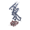
 Plasmodium falciparum erythrocyte membrane protein 1
  Keywords
 Function and homology information
 Authors
 Denmark, 2items
  Citation
 Structure visualization
 Downloads & links
 Other downloads

 PDBj
PDBj Assembly


 Sample preparation
 Electron microscopy imaging
 Processing