 ムービー
ムービー コントローラー


+ データを開く

- 基本情報
基本情報
| 登録情報 | データベース: PDB / ID: 8byg | |||||||||
|---|---|---|---|---|---|---|---|---|---|---|
| タイトル | fragment-linked stabilizer for ERa - 14-3-3 interaction (1047648) | |||||||||
 要素 要素 |
| |||||||||
 キーワード キーワード |  STRUCTURAL PROTEIN (タンパク質) / 14-3-3 (14-3-3タンパク質) / ERa / fragment linking / stabilization STRUCTURAL PROTEIN (タンパク質) / 14-3-3 (14-3-3タンパク質) / ERa / fragment linking / stabilization | |||||||||
| 機能・相同性 |  機能・相同性情報 機能・相同性情報regulation of epidermal cell division / protein kinase C inhibitor activity / positive regulation of epidermal cell differentiation / keratinocyte development / ケラチン / Regulation of localization of FOXO transcription factors / keratinocyte proliferation / phosphoserine residue binding / Activation of BAD and translocation to mitochondria / negative regulation of keratinocyte proliferation ...regulation of epidermal cell division / protein kinase C inhibitor activity / positive regulation of epidermal cell differentiation / keratinocyte development / ケラチン / Regulation of localization of FOXO transcription factors / keratinocyte proliferation / phosphoserine residue binding / Activation of BAD and translocation to mitochondria / negative regulation of keratinocyte proliferation / establishment of skin barrier / SARS-CoV-2 targets host intracellular signalling and regulatory pathways / Chk1/Chk2(Cds1) mediated inactivation of Cyclin B:Cdk1 complex / protein kinase A signaling / negative regulation of stem cell proliferation / SARS-CoV-1 targets host intracellular signalling and regulatory pathways / RHO GTPases activate PKNs / protein export from nucleus / negative regulation of innate immune response / protein sequestering activity / TP53 Regulates Transcription of Genes Involved in G2 Cell Cycle Arrest / release of cytochrome c from mitochondria / positive regulation of protein export from nucleus / stem cell proliferation / Translocation of SLC2A4 (GLUT4) to the plasma membrane / TP53 Regulates Metabolic Genes / negative regulation of protein kinase activity / negative regulation of cysteine-type endopeptidase activity involved in apoptotic process / intrinsic apoptotic signaling pathway in response to DNA damage / positive regulation of cell growth / regulation of cell cycle / cadherin binding / protein kinase binding / negative regulation of transcription by RNA polymerase II / シグナル伝達 / extracellular space / extracellular exosome / identical protein binding / 細胞核 / 細胞質基質 / 細胞質類似検索 - 分子機能 | |||||||||
| 生物種 |  Homo sapiens (ヒト) Homo sapiens (ヒト) | |||||||||
| 手法 | X線回折 / 分子置換 / 解像度: 1.7 Å | |||||||||
 データ登録者 データ登録者 | Visser, E.J. / Sijbesma, E. / Ottmann, C. | |||||||||
| 資金援助 |  オランダ, 1件 オランダ, 1件
| |||||||||
 引用 引用 | ジャーナル: Angew.Chem.Int.Ed.Engl. / 年: 2023 タイトル: From Tethered to Freestanding Stabilizers of 14-3-3 Protein-Protein Interactions through Fragment Linking. 著者: Visser, E.J. / Jaishankar, P. / Sijbesma, E. / Pennings, M.A.M. / Vandenboorn, E.M.F. / Guillory, X. / Neitz, R.J. / Morrow, J. / Dutta, S. / Renslo, A.R. / Brunsveld, L. / Arkin, M.R. / Ottmann, C. | |||||||||
| 履歴 |
|
- 構造の表示
構造の表示
| 構造ビューア | 分子: MolmilJmol/JSmol |
|---|
- ダウンロードとリンク
-ダウンロード
| PDBx/mmCIF形式 | 8byg.cif.gz | 112.3 KB | 表示 | PDBx/mmCIF形式 |
|---|---|---|---|---|
| PDB形式 | pdb8byg.ent.gz | 84.8 KB | 表示 | PDB形式 |
| PDBx/mmJSON形式 | 8byg.json.gz | ツリー表示 | PDBx/mmJSON形式 | |
| その他 |  その他のダウンロード その他のダウンロード |
-検証レポート
| アーカイブディレクトリ | https://data.pdbj.org/pub/pdb/validation_reports/by/8bygftp://data.pdbj.org/pub/pdb/validation_reports/by/8byg | HTTPS FTP |
|---|
-関連構造データ
| 関連構造データ | 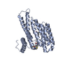 8bwjC 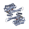 8bwxC 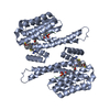 8bwzC  8bx0C 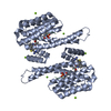 8bx3C  8bx4C  8bxiC  8bxmC  8bxnC  8bxoC 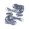 8bxqC 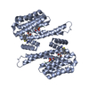 8bxsC  8by9C 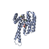 8bybC  8bycC 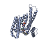 8bydC  8byeC  8byfC  8byoC  8byyC  8byzC 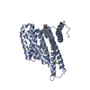 8bz0C 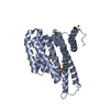 8bz9C 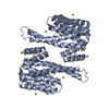 8bzaC  8bzbC  8bzwC  8c04C  8c0kC 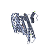 8c4fC 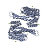 8c4gC C: 同じ文献を引用 ( |
|---|---|
| 類似構造データ |
-リンク
 PDBj
PDBj


- 集合体
集合体
| 登録構造単位 | 
| ||||||||
|---|---|---|---|---|---|---|---|---|---|
| 1 | 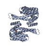
| ||||||||
| 単位格子 |
|
-要素
| #1: タンパク質 | 分子量: 26542.914 Da / 分子数: 1 / 由来タイプ: 組換発現 / 由来: (組換発現) Homo sapiens (ヒト) / 遺伝子: SFN, HME1 / 発現宿主:  Escherichia coli (大腸菌) / 参照: UniProt: P31947 Escherichia coli (大腸菌) / 参照: UniProt: P31947 |
|---|---|
| #2: タンパク質・ペプチド | 分子量: 613.596 Da / 分子数: 1 / 由来タイプ: 合成 / 由来: (合成) Homo sapiens (ヒト) |
| #3: 化合物 | ChemComp-S6O / ~{  分子量: 459.004 Da / 分子数: 1 / 由来タイプ: 合成 / 式: C23H27ClN4O2S 分子量: 459.004 Da / 分子数: 1 / 由来タイプ: 合成 / 式: C23H27ClN4O2S |
| #4: 水 | ChemComp-HOH / 水 分子量: 18.015 Da / 分子数: 335 / 由来タイプ: 天然 / 式: H2O 分子量: 18.015 Da / 分子数: 335 / 由来タイプ: 天然 / 式: H2O |
| 研究の焦点であるリガンドがあるか | Y |
-実験情報
-実験
| 実験 | 手法: X線回折 / 使用した結晶の数: 1 |
|---|
- 試料調製
試料調製
| 結晶 | マシュー密度: 2.67 Å3/Da / 溶媒含有率: 53.85 % |
|---|---|
| 結晶化 | 温度: 277 K / 手法: 蒸気拡散法, シッティングドロップ法 詳細: 0.095 M HEPES (pH 7.1), PEG400 (24% (v/v)), 0.19 M CaCl2 and 5% (v/v) Glycerol |
-データ収集
| 回折 | 平均測定温度: 100 K / Serial crystal experiment: N |
|---|---|
| 放射光源 | 由来: SEALED TUBE / タイプ: RIGAKU MICROMAX-003 / 波長: 1.54187 Å |
| 検出器 | タイプ: DECTRIS PILATUS 200K / 検出器: PIXEL / 日付: 2019年7月26日 |
| 放射 | プロトコル: SINGLE WAVELENGTH / 単色(M)・ラウエ(L): M / 散乱光タイプ: x-ray |
| 放射波長 | 波長: 1.54187 Å / 相対比: 1 |
| 反射 | 解像度: 1.7→66.36 Å / Num. obs: 189444 / % possible obs: 96.6 % / 冗長度: 6.1 % / CC1/2: 0.996 / Net I/σ(I): 11.6 |
| 反射 シェル | 解像度: 1.7→1.73 Å / Num. unique obs: 1263 / CC1/2: 0.488 |
- 解析
解析
| ソフトウェア |
| |||||||||||||||||||||||||||||||||||||||||||||||||||||||||||||||||||||||||||||||||||||||||||
|---|---|---|---|---|---|---|---|---|---|---|---|---|---|---|---|---|---|---|---|---|---|---|---|---|---|---|---|---|---|---|---|---|---|---|---|---|---|---|---|---|---|---|---|---|---|---|---|---|---|---|---|---|---|---|---|---|---|---|---|---|---|---|---|---|---|---|---|---|---|---|---|---|---|---|---|---|---|---|---|---|---|---|---|---|---|---|---|---|---|---|---|---|
| 精密化 | 構造決定の手法: 分子置換 / 解像度: 1.7→34.12 Å / SU ML: 0.2 / 交差検証法: FREE R-VALUE / σ(F): 1.34 / 位相誤差: 22.24 / 立体化学のターゲット値: ML
| |||||||||||||||||||||||||||||||||||||||||||||||||||||||||||||||||||||||||||||||||||||||||||
| 溶媒の処理 | 減衰半径: 0.9 Å / VDWプローブ半径: 1.11 Å / 溶媒モデル: FLAT BULK SOLVENT MODEL | |||||||||||||||||||||||||||||||||||||||||||||||||||||||||||||||||||||||||||||||||||||||||||
| 精密化ステップ | サイクル: LAST / 解像度: 1.7→34.12 Å
| |||||||||||||||||||||||||||||||||||||||||||||||||||||||||||||||||||||||||||||||||||||||||||
| 拘束条件 |
| |||||||||||||||||||||||||||||||||||||||||||||||||||||||||||||||||||||||||||||||||||||||||||
| LS精密化 シェル |
|
